1. Treatment
1.1. -Anticoagulant:( Heparin, warfarin) to prevent new clots from forming. -Clot dissolvers (thrombolytics): These drugs speed up the breakdown of a clot. -Surgery may be necessary to remove problematic clots, especially those that restrict blood flow to the lungs or heart.
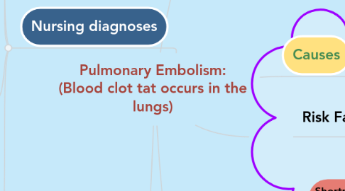
2. Nursing diagnoses
2.1. Impaired gas exchanged related to decrease pulmonary perfusion associated with obstruction of pulmonary arterial blood flow by the embolus.
2.1.1. Nursing intervention:1-Frequently assess respiratory status including rate, depth, effort, lung sound and SPO2.2-Assess the mental status of the client.3-Monitor ABGs and note changes.4-Position the patient in high Fowler¶position.5-Administered oxygen as ordered by doctor.6-Maintain bed rest.7-Administer medications(anticoagulants) as prescribed by doctor.
2.2. Ineffective Breathing Pattern related to hypoxia,anxiety and fatigue.
2.2.1. 1-Monitor oxygen saturation as indicated.2-Assess the characteristics of pain, especially in association with the respiratory cycle.3-Assess the client’s anxiety level.4-Administer oxygen as indicated.5-Anticipate the need for intubation and mechanical ventilation.
2.3. Deficient Knowledge related to new medical condition and new treatment
2.3.1. 1-Assess the client’s knowledge of pulmonary embolus: its severity, prognosis, risk factors, and therapy.2-Provide information.3-Inform the client of the need for routine laboratory testing while on oral anti coagulation.4-Discuss and give the client a list of signs and symptoms of excessive anti coagulation.5-Explain the need for a vena cava filter device if clotting is a chronic problem.
2.4. Risk for Bleeding related to anticoagulant or thrombolytic therapy.
2.4.1. 1-Assess for the signs and symptoms of bleeding.2-Assess for the history of a high-risk bleeding condition.3-Monitor platelet counts, coagulation test results.4-If bleeding occurs while on heparin, anticipate the following: Stop the infusion Recheck the aPTT level stat Take vital signs often Reevaluate the dose of heparin on the basis of the aPTT result Notify the blood bank to ensure blood availability if needed.
