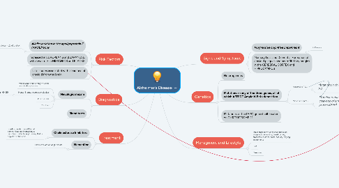
1. Risk Factors
1.1. Allelic variation in the apolipoprotein E (APOE) locus
1.1.1. 3 major alleles = e2, e3 and e4
1.1.1.1. individuals with one copy of the e4 allele are 2- to 5 TIMES MORE LIKELY to develop AD
1.1.1.1.1. two copies = 5 to 10 TIMES MORE LIKELY
1.2. Increased in EUROPEANS and JAPANESE, with lower risk in HISPANICS and BLACKS.
1.3. Late stages associated with clearance of amyloid from the brain
2. Diagnostics
2.1. Neuological exam
2.1.1. reflexes, tone and strength, vision, coordination, balance
2.1.2. Mental Status, neuropsychological
2.1.2.1. Mini Mental State Exam (MMSE)
2.1.3. MRI, CT, PET
2.1.4. CSF fluid
2.2. Blood tests
3. Treatment
3.1. Cholinesterase Inhibitors
3.1.1. boosting levels of a cell-to-cell communication by providing a neurotransmitter (acetylcholine) that is depleted in the brain
3.2. Memantine
3.2.1. Blocks the action of glutamate
4. Signs and Symptoms
4.1. Progressive cognitive impairment
4.1.1. 7-10 years
4.2. Memory Loss and Dementia, Formation of amyloid plaques and neurofibrillary tangles in the CEREBRAL CORTEX and HIPPOCAMPUS
5. Genetics
5.1. Heterogenous
5.2. Mutations in any of the three genes, all of which AFFECT Amyloid-Beta deposition
5.2.1. Presenilin 1 (PS1)
5.2.1.1. Mutations in this gene result in early onset AD
5.2.2. Presenilin 2 (PS2)
5.2.2.1. Protein products are involved in the cleavage of amyloid-B precursor protein (APP)
5.2.2.1.1. When (APP) is not CLEAVED normally, accumulates excessively and is DEPOSITED in the brain.

