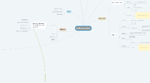
1. Observation
1.1. General
1.1.1. Color
1.1.2. Edema
1.1.3. Wounds
1.1.4. Deformity
1.2. Lateral/Medial Malleolus
1.2.1. Swelling?
1.2.2. Ecchymosis?
1.3. Sinus Tarsi
1.3.1. Concave shape (swollen)
1.3.2. Girth Measurement
1.3.2.1. figure-8
1.3.2.2. "zero point" at ant. edge of med or lat malleolus
1.3.2.3. perform bilat
1.3.2.4. (+) test: sig. difference bilat
1.4. Achilles Tendon
1.4.1. Thickening
2. Palpation
2.1. Tenderness?
2.1.1. Ottawa Ankle Rules: if (+) refer for x-ray (pg. 279)
2.1.1.1. Malleolar zone
2.1.1.2. Apex of Lateral Malleolus
2.1.1.3. Post.Lat Malleolus
2.1.1.4. Medial Malleolus
2.1.1.5. unable to bear normal weight
2.2. Divots?
2.3. Deformities?
2.4. Crepidus?
3. AROM/PROM (endfeel!)
3.1. Inversion/Eversion
3.1.1. Fulcrum: Over talus in center of malleolus Stationary: Midline of tibia Movement: Midline of 2nd metatarsal
3.1.1.1. 25 degrees of ROM
3.1.1.1.1. Inversion
3.1.1.1.2. Eversion
3.2. Plantar/Dorsiflexion
3.2.1. Fulcrum: Center of Lat. Malleolus Stationary: Long axis of Fibula Movement: Parallel w/ bottom of foot
3.2.1.1. 70 degrees ROM
3.2.1.1.1. Dorsiflexion
3.2.1.1.2. Plantarflexion
4. Neuro
4.1. Lower Quarter Screen
4.2. Common Peroneal Nerve
4.3. Tibial Nerve
5. Diagnosis
5.1. Lateral Ankle Sprain
5.1.1. History
5.1.1.1. acute
5.1.1.2. pain
5.1.1.2.1. lat. malleolus/sinus tarsi
5.1.1.3. Pt may report "pop"
5.1.1.4. mechanism
5.1.1.4.1. supination
5.1.1.4.2. plantar flexion
5.1.2. Func. Assessment
5.1.2.1. gait w/ shortened stance
5.1.2.2. inability to stand on injured leg
5.1.3. Observation
5.1.3.1. swelling
5.1.3.1.1. lat. joint capsul
5.1.3.2. eechymosis
5.1.3.2.1. lat. malleolus
5.1.4. Palpation
5.1.4.1. pain
5.1.4.1.1. along lig.
5.1.4.2. crepitus
5.1.4.2.1. lig. origin or insertion
5.1.5. AROM/PROM
5.1.5.1. pain on lat side
5.1.5.1.1. stretching of lat ligs
5.1.5.2. pain on med side
5.1.5.2.1. pinching of med structures
5.1.6. MMT
5.1.6.1. peroneals weak
5.1.6.2. extensors weak
5.2. High Ankle Sprain
5.2.1. History
5.2.1.1. acute
5.2.1.2. pain
5.2.1.2.1. ant. distal tib. fib.
5.2.1.3. mechanism
5.2.1.3.1. int. rotation of talus
5.2.1.3.2. ext. rotation of talus
5.2.2. Func Assessment
5.2.2.1. gait shortened
5.2.2.2. decreased strength w/ push off
5.2.3. Observation
5.2.3.1. swelling
5.2.3.1.1. distal tib fib
5.2.4. Palpation
5.2.4.1. pain
5.2.4.1.1. distal tib fib
5.2.4.2. fibula
5.2.4.2.1. rule out fx
5.2.5. AROM/PROM
5.2.5.1. motion is restricted
5.2.5.1.1. especially w/ dorsiflexion and eversion
5.2.6. MMT
5.2.6.1. tibialis ant. and post. weak and painful
5.3. Medial Ankle Sprain
5.3.1. History
5.3.1.1. acute
5.3.1.2. pain
5.3.1.2.1. med. boarder of ankle/foot radiating from med. malleolus
5.3.1.3. mechanism
5.3.1.3.1. eversion/rotation
5.3.2. Func Assessment
5.3.2.1. gait shortened/painful midstance
5.3.3. Observation
5.3.3.1. swelling
5.3.3.1.1. med. joint capsul
5.3.4. Palpation
5.3.4.1. pain
5.3.4.1.1. deltoid ligs
5.3.4.2. crepitus
5.3.4.2.1. lig origin/inserrtion
5.3.5. AROM/PROM
5.3.5.1. pain med. side during dorsiflexion
5.3.5.1.1. stretching ant tib talar/navic
5.3.5.2. pain during dorsiflexion
5.3.5.2.1. trauma to post tib talar
5.3.5.3. lat pain
5.3.5.3.1. pinching/trauma to lat side
5.3.6. MMT
5.3.6.1. post tibialis weak/painful
5.4. Achilles Tendon Rupture
5.4.1. History
5.4.1.1. acute
5.4.1.2. pain
5.4.1.2.1. achilles tendon/back of gastro
5.4.1.3. Pt reports "pop"
5.4.1.4. mechanism
5.4.1.4.1. forceful dorsi/plantar flexion
5.4.1.4.2. eccentric loading/plyometric contraction of calf
5.4.2. Func Assessment
5.4.2.1. unable to perform heal raise
5.4.2.2. unable to push off
5.4.3. Observation
5.4.3.1. defect may be visable
5.4.3.2. rapid swelling
5.4.3.3. discoloration
5.4.4. Palpation
5.4.4.1. defect in achilles tendon
5.4.4.2. pain along tendon/lower gastroc
5.4.5. AROM/PROM
5.4.5.1. plantarflexion may be possible
5.4.5.2. pain w/ dorsiflexion
5.4.6. MMT
5.4.6.1. weak/absent plantarflexion
5.5. Subluxation
5.5.1. History
5.5.1.1. acute or insideous
5.5.1.2. pain
5.5.1.2.1. behind lat mllelous
5.5.1.2.2. Pt reports instability and "snapping"
5.5.1.2.3. Mechanism
5.5.2. Func Assessment
5.5.2.1. reproduce symptoms w/ rapid direction changes
5.5.3. Observation
5.5.3.1. swelling and ecchymosis
5.5.4. Palpation
5.5.4.1. tender behind lat mallelous
5.5.5. AROM/PROM
5.5.5.1. pereneal tendon mvmt
5.5.6. MMT
5.5.6.1. pereneals
5.6. Med/Tib Stress Syndrom
5.6.1. History
5.6.1.1. insideous
5.6.1.2. pain
5.6.1.2.1. tibia
5.6.1.3. mechanism
5.6.1.3.1. overuse stress
5.6.2. Func Asessment
5.6.2.1. gait w/ excessive pronation
5.6.3. Observation
5.6.3.1. Pes Planus
5.6.3.2. rear/forefoot varus in STJN
5.6.4. Palpation
5.6.4.1. tender along tibia
5.6.5. AROM/PROM
5.6.5.1. increase pain w/ dorsiflexion, pronation, toe extension
5.6.6. MMT
5.6.6.1. symptoms reproduced w/ repetitions of muscles used
5.7. Stress Fracture
5.7.1. History
5.7.1.1. chronic or insideous
5.7.1.2. pain
5.7.1.2.1. shaft of tib/fib
5.7.1.2.2. localized during activity
5.7.1.2.3. achy while at rest
5.7.1.3. mechanism
5.7.1.3.1. sudden increase in duration, frequency, intensity or change in surface or footwear
5.7.2. Func Assessment
5.7.2.1. shortened stance phase
5.7.3. Observation
5.7.3.1. localized swelling possible
5.7.4. Palpation
5.7.4.1. pain along fx site
5.7.5. AROM/PROM
5.7.5.1. n/a
5.7.6. MMT
5.7.6.1. decreased strength of muscles near fx
5.8. Fracture
5.8.1. History
5.8.1.1. acute
5.8.1.2. pain
5.8.1.2.1. sharp
5.8.1.2.2. localized
5.8.1.3. mechanism
5.8.1.3.1. direct blow
5.8.1.3.2. eversion/inversion stress
5.8.2. Func Assessment
5.8.2.1. n/a
5.8.3. Observation
5.8.3.1. swellin/ecchymosis may be prersent
5.8.3.2. deformity may be present
5.8.4. Palpation
5.8.4.1. point tender
5.8.4.2. possible crepitus
5.8.4.3. possible deformity
5.8.5. AROM/PROM
5.8.5.1. do NOT do with obvi fx
5.8.6. Tests
5.8.6.1. Squeeze test
5.8.6.1.1. do NOT do with obvi fx
6. History
6.1. Who are you?/What do you play?
6.2. What happened/MOI?
6.2.1. Inversion?
6.2.2. Eversion?
6.2.3. Plantarflexion?
6.2.4. Dorsiflexion?
6.3. When did this happen?
6.4. Where is your pain located?
6.5. Has this/similar happened before?
6.6. What type of pain do you feel?
6.7. What have you done since the injury?
6.8. Has your level of activity increased or decreased?
6.9. Did you hear/feel anything?
7. MMT
7.1. Dorsifelxion and Supination
7.1.1. Pt. position
7.1.1.1. seated
7.1.2. Test position
7.1.2.1. knee flexed
7.1.2.2. foot dorsiflexed and supinated
7.1.3. Stabilization
7.1.3.1. distal tibia
7.1.4. Resistence
7.1.4.1. top of the foot
7.2. Eversion and Pronation
7.2.1. Pt position
7.2.1.1. laying on non-injured side
7.2.1.2. non-injured hip is flexed
7.2.2. test position
7.2.2.1. test foot off end of table
7.2.3. stabilization
7.2.3.1. lower leg
7.2.4. resistance
7.2.4.1. lateral side of foot
7.3. Plantar Flexion
7.3.1. Pt position
7.3.1.1. prone
7.3.2. test position
7.3.2.1. Gastrocnemius: straight leg, foot off table
7.3.2.2. Soleus: knee bent past 3o degrees
7.3.3. stabilization
7.3.3.1. prox. to ankle
7.3.4. resistence
7.3.4.1. bottom of foot
7.4. Rearfoot Inversion
7.4.1. Pt position
7.4.1.1. laying on injured side
7.4.1.2. other hip flexed
7.4.2. test position
7.4.2.1. test foot off table
7.4.3. stabilization
7.4.3.1. med. aspect of lower leg
7.4.4. resistance
7.4.4.1. middle of foot
8. Stress Tests
8.1. Ant. Drawer Test
8.1.1. Pt. Position
8.1.1.1. Sitting
8.1.1.2. knee flexed
8.1.2. ATs Position
8.1.2.1. sitting in front of Pt
8.1.2.2. hand placement
8.1.2.2.1. stabilize: lower leg/right above ankle
8.1.2.2.2. cup calc. w/ foot resting on forearm
8.1.3. Procedure
8.1.3.1. calc. drawn fwd towards AT
8.1.4. (+) Test
8.1.4.1. pain
8.1.4.2. talus slides towards AT
8.1.4.3. "clunk"
8.1.5. Implications
8.1.5.1. anterior talofibular sprain
8.2. Talar Tilt
8.2.1. Pt Position
8.2.1.1. sitting w/ legs over edge of table
8.2.2. ATs Position
8.2.2.1. in front of Pt
8.2.2.2. hand placement
8.2.2.2.1. stabilize:thumb or finger on calcaneofibular ligament to feel
8.2.2.2.2. grasp calc. and talus
8.2.3. Procedure
8.2.3.1. roll calc. medially and laterally, causing talus to tilt
8.2.4. (+) Test
8.2.4.1. bilat difference
8.2.4.2. pain
8.2.5. Implications
8.2.5.1. Inversion
8.2.5.1.1. involvement of of ligaments
8.2.5.2. Eversion
8.2.5.2.1. deltoid lig. sprain

