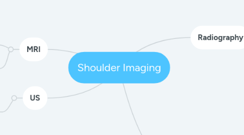
Shoulder Imaging
by Heather Berge


1. Radiography
1.1. AP internal rotation: shows the lesser tuberosity in profile, medially, and the greater tuberosity projected over the center of the humeral head.
1.2. AP external rotation: shows the greater tuberosity in profile, laterally, and the lesser tuberosity over the center of the head.
1.3. Axillary: true lateral of the shoulder joint, able to visualize anterior or posterior dislocations.
1.4. Transcapular Y: true lateral of the scapula that shows the position of the humeral head within the joint. It can be used to show anterior vs. posterior dislocations.
1.5. Grashey: true AP of the shoulder joint with the glenohumeral joint space open and the glenoid fossa in profile.
1.6. Outlet view: Transcapular Y view with a 10 degree caudal angle. Shows the underside of the acromion process and is useful for evaluation of rotator cuff impingement.
2. CT
2.1. CT Arthrography: contrast injected into the joint capsule prior to scan.
2.1.1. Shows labral tears, the joint capsule, rotator cuff tears, and articular cartilage defects.
2.2. CT without contrast
2.2.1. Shows dislocations, fractures, soft-tissue calcifications, and the glenoid and humeral head morphology.
3. MRI
3.1. MRI Arthrography: the gold standard for evaluating the labrum
3.1.1. May be done with gadolinium or saline injected into the joint space.
3.2. MRI without contrast
3.2.1. Planes scanned in oblique coronal and oblique sagittal (parallel to the rotator cuff and glenoid articular surface, respectively)

