
Fetal Monitoring
by Toni Zappulla
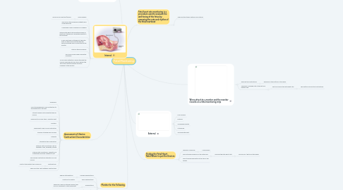
1. Patient must have some dilation to the cervix to apply
2. Internal
2.1. More invasive
2.1.1. More risk for infection/trauma
2.2. Involves a uterine pressure catheter and scalp electrode
2.3. Used when closer surveillance is needed
2.4. Internal fetal heart rate monitoring uses an electronic transducer connected directly to the fetal skin
2.5. A wire electrode is attached to the fetal scalp or other body part through the cervical opening and is connected to the monitor
2.6. Internal uterine pressure
2.7. Will only be done when absolutely necessary
2.8. A fluid-filled catheter is placed through the cervical opening into the uterus beside the fetus and transmits uterine pressure readings to the monitor
3. Assessment of Uterine Contraction Characteristics
3.1. Frequency
3.2. From the beginning of one contraction to the beginning of the next
3.3. Intensity should not be greater than 90 mmHG
3.4. Should not be closer than 2 minutes apart
3.5. Duration
3.6. Beginning to end of one contraction
3.7. Should not exceed 90 seconds
3.8. Intensity
3.9. Strength of the contraction
3.10. External Fetal Monitoring- felt by palpation: mild, moderate, strong
3.11. Internal Fetal Monitoring : consists of a intrauterine pressure catheter(IUCP)
3.12. The average contraction intensity is 50-85 mmHG
3.13. Resting tone:
3.13.1. Must not be greater than 20mmHG
3.14. There must be “rest” between contractions
4. Monitor for the Following
4.1. Variable decelerations
4.1.1. Require interventions
4.2. Early decelerations
4.2.1. Continue to monitor
4.3. Accelerations
4.3.1. These are a sign of fetal well-being, they occur in response to fetal movement
4.4. Late decelerations
4.4.1. Require interventions
4.5. Prolonged decelerations
4.5.1. Require interventions
5. Wires attach to a monitor and the monitor records on a fetal monitoring strip
5.1. Records the contractions
5.1.1. Records on the bottom of the paper
5.2. The paper is divided into a top part and bottom part
5.2.1. The top records the fetal heart rate
5.2.1.1. The bottom records the contractions
6. Fetal heart rate monitoring is a procedure used to evaluate the well-being of the fetus by assessing the rate and rhythm of the fetal heartbeat
6.1. There are two types: External and Internal
7. External
7.1. Less invasive
7.2. External
7.3. Tocodynamometer
7.4. Ultrasound
7.5. Secured with belts
8. Finding the Fetal Heart Rate/Where to put the Devices
8.1. Leopold's Maneuver
8.1.1. 4 manuevers
8.2. Place ultrasound device on the fetal back
8.2.1. Records the fetal heart rate
8.2.1.1. Records on the top of the paper