1. Gain in attachment: Repair, Regeneration
2. Radiographs: Absence doesn't mean calculus isn't present.
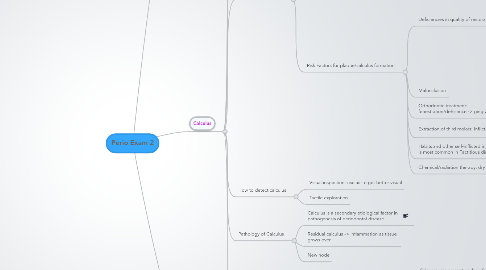
3. Calculus
3.1. What is Calculus?
3.1.1. Mineralized bacterial plaque that forms on surfaces of teeth and dental prosthesis.
3.2. Types of Calculus
3.2.1. Supragingival
3.2.1.1. q Coronal to gingival margin q Visible q White or whitish yellow q Clay-‐like consistency q Easily detached q Saliva-‐Mineralization
3.2.2. Subgingival
3.2.2.1. q Below the gingival margin q Not visible q Dark brown/ greenish black q Hard and dense q Firmly attached to tooth q Gcf
3.2.2.2. Subgingival is harder than supragingival
3.3. Attachment
3.3.1. Attaches firmly to non-shedding surfaces, especially rough/grooved.
3.3.2. Root planning smooths the root surface -> reduces calculus adherence.
3.4. Composition of Calculus
3.4.1. Inorganic 70-90%
3.4.1.1. 76% Calcium Phosphate
3.4.1.2. 66% is in crystalline form
3.4.1.3. Major crystalline form: Hydroxyapatite 58%
3.4.2. Organic 10-30%
3.5. Distribution
3.5.1. Supragingival: Hydroxyapatite 58%
3.5.2. Subgingival: Hyroxyapatite, whitlockite
3.5.3. Mandibular anterior region: Brushite
3.5.4. Posterior areas: Magnesium whitlockite
3.6. Formation of calculus
3.6.1. Acquired pellicle: within minutes
3.6.2. Plaque Maturation: 24-48 hrs
3.6.3. Plaque mineralization: 1-14 days of plaque formation
3.6.4. Theories regarding mineralization
3.6.4.1. Rise in Ph of saliva: Ammonia is produced by bacteria
3.6.4.2. Colloidal proteins in saliva bind calcium/phosphate salts->Supersaturation.
3.6.4.3. Phosphatase from dental plaque
3.6.4.4. Most widely accepted: Epitatic concept/Heterogenous nucleation
3.6.4.4.1. Focal seeding points attract minerals-> expansion.
3.6.4.4.2. Seeding agent: Charge of Glycocalyx??
3.6.5. Risk Factors for plaque/calculus formation
3.6.5.1. Deficiencies in quality of restoration
3.6.5.1.1. Margin of restoration
3.6.5.1.2. Contours/open contacts
3.6.5.1.3. Materials and design
3.6.5.1.4. Roughness
3.6.5.2. Malocclusion
3.6.5.3. Orthodontic treatment: fenestration/dehisence -> gingival recession
3.6.5.4. Extraction of third molars: Inflict bone defects
3.6.5.5. Habits and other self-inflicted injures: Canine is most common in Factitious disorders
3.6.5.5.1. Treat this disorder before perio surgery
3.6.5.6. Chemical/radiation therapy: dry mouth
3.7. How to detect calculus
3.7.1. Visual inspection: use air to get better visual
3.7.2. Tactile exploration
3.8. Pathology of Calculus
3.8.1. Calculus is a secondary etiological factor in pathogenesis of periodontal disease.
3.8.2. Residual calculus -> Inflammation as tissue grows over.
3.8.3. New node
3.9. Clinical Application
3.9.1. Supragingival calculus visible 2wks post SRP
3.9.1.1. Calculus was present under inflamed tissue
3.9.1.2. After 1st tx gingiva reduced -- This is not recession
3.9.1.3. Check previous quadrants when doing SRP
3.9.2. Reduction of probing depth 6wks post SRP
3.9.2.1. 1. Reduction of inflammation --> reduction of probing depth. This isn't healing.
3.9.2.2. 2. Healing: epithelial cells at junctional epithelium proliferate-->formation of long junctional epithelium. This is repair.
3.9.2.3. 3. Also gain attachment with increased Epithelium, CT, and bone. This is regeneration. Can only be determined in biopsy.
3.9.3. Calculus and salivary ducts
3.9.3.1. Wortons: Submandibular gland
3.9.3.2. Upper 1st molar -- Stenson: Parotid
3.9.3.3. Lower Incisors -- ?: Sublingual
3.9.4. General Prevention of calculus accumulation
3.9.4.1. Personal plaque control
3.9.4.2. Professional removal of calculus
3.9.5. What's wrong in these images?
3.9.5.1. Biologic Width violation is most likely. Maybe left over cord.
3.9.5.2. Most likely inflammation from plaque. Maybe hyperproliferation or early eruption.
3.9.5.3. Facitious disorder
3.9.5.4. Resorption of bone. Primary etiological factor is piercing. Plaque is secondary (unless plaque is present elsewhere).
