
ECG - Ventricular
da Naomi Rumrill
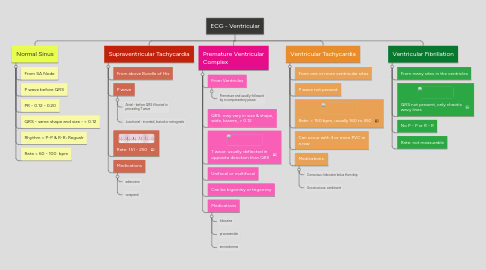
1. Normal Sinus
1.1. From SA Node
1.2. P wave before QRS
1.3. PR - 0.12 - 0.20
1.4. QRS - same shape and size - < 0.12
1.5. Rhythm = P-P & R-R; Regualr
1.6. Rate = 60 - 100 bpm
2. Supraventricular Tachycardia
2.1. From above Bundle of His
2.2. P wave
2.2.1. Atrial - before QRS if buried in preceding T wave
2.2.2. Junctional - inverted, buried or retrograde
2.3. Rate: 151 - 250
2.4. Medications
2.4.1. adenosine
2.4.2. verapamil
3. Premature Ventricular Complex
3.1. From Ventricles
3.1.1. Premature and usually followed by a compensatory pause
3.2. QRS: may vary in size & shape, wide, bizarre, > 0.12
3.3. T wave: usually deflected in opposite direction than QRS
3.4. Unifocal or multifocal
3.5. Can be bigeminy or trigeminy
3.6. Medications
3.6.1. lidocaine
3.6.2. procanamide
3.6.3. amniodorone
4. Ventricular Tachycardia
4.1. From one or more ventricular sites
4.2. P wave not present
4.3. Rate: < 150 bpm, usually 160 to 350
4.4. Can occur with 3 or more PVC in a row
4.5. Medications:
4.5.1. Conscious: lidocaine bolus then drip
4.5.2. Unconscious: cardiovert