
ECG - Blocks
作者:Naomi Rumrill
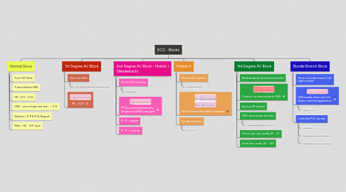
1. Normal Sinus
1.1. From SA Node
1.2. P wave before QRS
1.3. PR - 0.12 - 0.20
1.4. QRS - same shape and size - < 0.12
1.5. Rhythm = P-P & R-R; Regualr
1.6. Rate = 60 - 100 bpm
2. 1st Degree AV Block
2.1. From the Atria
2.1.1. Delay between the atria and bundle of his
2.2. PR > 0.20
3. 2nd Degree AV Block - Mobitz I (Wenkebach)
3.1. Block at AV junction
3.1.1. progressive
3.2. PR becomes progressively longer until QRS is dropped
3.3. P - P = regular
3.4. R - R = irregular
4. Mobitz II
4.1. Block at AV junction
4.1.1. intermittent block
4.2. More P waves then QRS complexes
4.3. Usually a pattern
4.3.1. 2:1, 3:1, 4:1
5. 3rd Degree AV Block
5.1. Block between atria and ventricles
5.2. P waves: no relationship to QRS
5.3. No true PR interval
5.4. QRS: same shape and size
5.4.1. Usually wide, bizarre and > 0.12
5.5. Ventricular rate usually 20 - 40
5.6. Atrial rate usually 60 - 100
6. Bundle Branch Block
6.1. Block in bundle branch (left, right or both)
6.2. QRS usually same size and shape, notched appearance
6.2.1. Usually > 0.12
6.3. Looks like PVC except:
6.3.1. Has P wave
6.3.2. t wave going in same direction
6.3.3. Initiated by SA node, not ventricle