

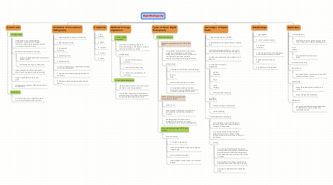
1. General Info
1.1. 1- Digital image:
1.1.1. image saved in a computer memory composed of discrete units of digital information called pixels arranged in the form of a matrix of rows and columns
1.1.2. are numeric and discrete in two ways
1.1.2.1. in terms of spatial distribution of the picture elements pixels
1.1.2.2. different shades of gray of each pixel
1.1.3. image represent the different penetration value of each x ray photon through the body
1.1.4. image is presented by 20-30 micron pixels
1.1.5. digital matrix composed of 256 shades 0 black and 256 white
1.2. Digitialization
1.2.1. convert an image into a digital form that in turn can be processed by the computer
2. Compnents
2.1. 1. xray machine
2.2. 2. sensor
2.3. 3. computer
2.4. 4. monitor
2.5. 5. printer
3. Methods of image digitization:
3.1. 1- indirect digital radiography
3.1.1. procedure of transferring chemically processed analogue ( film based 0 image to computer by scanner or camera
3.1.2. disadvantages
3.1.2.1. 1. doesn't solve processing problems
3.1.2.2. 2. time and effort consuming
3.1.2.3. 3. result in loss and alteration of information
3.2. 2- Direct digital radiography
3.2.1. image receptor instead of x ray film is called the sensor or the image plate (ip)
3.2.2. xray reaching image plate is converted into electric energy to be analyzed and digitized by computer producing digital image
4. Types od Direct Digital Radiography
4.1. 1- Solid state detectors
4.1.1. charged coupled dentistry CCD (RVG) Real Time
4.1.1.1. x ray photons reaching sensor are converted to light by an intensifying or scintillation screen, then it is picked by ccd and converted into electrical charge, then relayed to computer producing digital image on monitor
4.1.1.2. disadvantages
4.1.1.2.1. active area is smaller than total surface area
4.1.1.2.2. sensor is bulky
4.1.1.2.3. electronic cable annoying to patient
4.1.1.2.4. some replaced caple by microwave transmitter but needs additional electornic component increasing bulk of sensor
4.1.2. CMOS complementary metal oxide semiconductors ) (RVG)
4.1.2.1. similar to ccd
4.1.2.2. differ that each CMOS pixel is isolated from each neighbor and directly connected to transistor
4.1.2.3. the charge packet from each pixel is transferred to the transistor as voltage anabling each pixel to be assessed seperately
4.1.3. photo stimulable phosphor PSP (Digora) cordless
4.1.3.1. process occurs by
4.1.3.1.1. 1- storage of xray energy
4.1.3.1.2. read out by helium neon laser which releases image as light
4.1.3.1.3. photo multiplier detect light
4.1.3.1.4. analog digital converter convert it to electrical charges
5. Advantages of Digital Radio
5.1. 1. reduced exposure time (60-008)
5.2. 2. standardization and reproductibilty in research work
5.3. 3. effective patient education tool : can view it with operator facilitating dialogue and rapport
5.4. 4. better case presentation, demonstration, and education
5.5. 5. image enhancment and manipulation
5.5.1. contrast brightness
5.5.2. zoom
5.5.3. mirror imaging
5.5.4. black and white conversion
5.6. 6. software advantages
5.6.1. distance and angle measurement
5.6.2. implant planning
5.7. 7. digital subtraction radiography
5.7.1. permit operator to remove all anatomic structures that haven't changed between radiographic examination
5.7.2. for easy identification of any changes in diagnostic information ( change in bone density, dimension that may occur follow up of pdl disease
5.7.3. method
5.7.3.1. 2 or more reproducible digital images are similarly made for the patient, first called inital base line image before dental therapy begin, 2nd is taken after period of time called follow up image
5.7.3.2. both introduced to software where both are superimposed on each other and compared
5.7.3.3. new image is generated called subtracted image
6. Disadvantage
6.1. 1. high expense
6.2. 2. costly ro replace receptor
6.3. 3. resolution limitatioon
6.4. 4. film bending to accommodate patient anatomy induce artifacts in receptor
6.5. 5. patient discomfort due to bulk
6.6. 6. receptors can't be sterilized
7. Application
7.1. 1- caries diagnosis
7.1.1. automated caries recognition program which look for reduction of density indicating caries
7.2. 2- pdl disease
7.2.1. loss of alveolar bone density or height
7.3. 3- periapical pathology
7.4. bone disease
7.4.1. early identification of osteo porosis and other metabolic disease of bone
7.5. implantology
7.5.1. assess bone quantity and quality prior to implant
7.5.2. assess changes after placement
7.6. orthodontics
7.6.1. consistent automatic landmarks identification of cephalometric images followed by craniofacial analysis