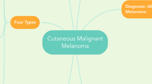
1. Four Types
1.1. Lentigo Maligna Melanoma
1.1.1. Shades of pigmentation/ irregular margins
1.1.2. Dark/ black-blue nodules
1.1.3. Atypical melanocytes singly in nests
1.1.4. Usually occurs in elderly
1.2. Acral Lentiginous Melanoma
1.2.1. Occurs hands/ feet/ sublingual areas
1.2.2. Atypical melanocytes of epidermal hyperplasia w/ rete ridges
1.3. Superficial Spreading Melanoma
1.3.1. Occurs young adults and adults mainly caucasian
1.3.2. Common with trunk and extremities
1.3.3. Papules/ nodules/ diffuse induration
1.3.4. Black/ blue/ tan/ pink colored
1.3.5. "Histopathologic findings include large melanocytes with dark, atypical nuclei and abundant, pale cytoplasm similar to the epidermal changes in Paget's disease" (Su 1997, p. 270).
1.4. Nodular Melanoma
1.4.1. Deep pigmentation w/ rapid growth patterns and ulcerations
1.4.2. Massive tumor growth in dermis
1.4.3. Atypical melanocytes of epidermis in nests or pagetoid pattern
2. Pathophysiology etiology
2.1. "Melanomas arise as a result of malignant degeneration of melanocytes located either along the basal layer of the epidermis or in benign melanocytes nevus" (Huether & McCance 2014, p. 1643).
2.2. Originates in melanocytes
2.3. MC1R genetic polymorphism
2.4. "Inactivating mutations in the CDKN2A gene which encodes for p16INK4a tumor suppressor protein, pose a high risk for development of cutaneous melanoma. Both BRAF and CDKN2A mutations in cutaneous melanoma cells are characteristic of indirect UV-induced oxidative damage" (Feller, Khammisa, Kramer, Altini & Lemmer 2016, p. 5).
3. Treatments
3.1. Interferon alpha-2b
3.2. Vemurafenib
3.3. Ipilimumab
3.4. Biopsy and removal
3.5. Radiation and chemoactive agents
3.6. High-dose interleukin-2
3.7. Adoptive T-cell therapy
4. At Risk Patients
4.1. Personal history
4.1.1. Sunlight exposure
4.1.1.1. Ultra violent B
4.1.1.2. Ultra violent A
4.1.2. History of sunburns
4.1.2.1. At young ages
4.1.2.2. Double the risk
4.1.3. Associated diseases
4.1.3.1. Pancreatic cancer
4.1.3.2. Retinoblastoma
4.1.3.3. Li-Fraumeni
4.1.3.4. Xeroderma Pigmentosa
4.2. Phenotype features
4.2.1. Fair complexion
4.2.2. Blue/ green eye color
4.2.3. Light/ red hair color
4.2.4. Presence of freckles
4.2.5. Nevi and location/ number
4.2.6. Inability to tan
4.3. Family history
4.3.1. "Person's with a first-degree relative with melanoma is one of the strongest predictive factors for the development of the disease" (Springer & McClary 2013, p. 40).
5. Facts
5.1. Horizontal vs vertical growth
5.1.1. "Metastatic involvement does not pose a threat in the horizontal growth phase because blood vessels do not occur in the epidermis" (Su 1997, p. 267).
5.1.2. Vertical growth can cause malignancies due to growth vertically into the dermis of the skin invading blood vessels and surrounding tissues.
