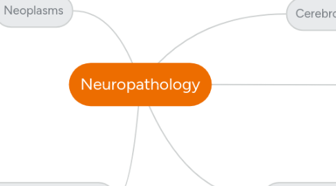
1. Cerebrovascular Disease
1.1. Ischemia
1.1.1. Ischemia and infarction in the brain have several causes. Ischemia may be divided into two types, global and focal.
1.1.1.1. Global
1.1.1.1.1. Global ischemia results from a generalized decrease in perfusion. Causes of global ischemia include shock/hypotension and cardiac arrest.
1.1.1.2. Focal
1.1.1.2.1. Focal ischemia results from decreased perfusion of a certain area of the brain. Causes of focal ischemia include emboli, thrombosis, and vasculitis.
1.2. Infarction
1.2.1. Infarcts occur with sustained reduced perfusion. Infarcts may be hemorrhagic or nonhemorrhagic. Hemorrhagic infarcts may occur when there is reperfusion of an infarcted segment of brain.
1.2.2. Histologically, the appearance of an infarct changes over time. After about 12 hours, ischemic change can be detected as eosinophilic change in neurons (“red neurons”), edema, and swelling of glial cells. At about 48 hours, there is an infiltrate of neutrophils and macrophages. Macrophages contain lipid (myelin) and may remain at the site of infarction for months. Reactive, enlarged glial cells with enlarged nuclei may be seen after about one week, persisting for several months.
1.3. NEUROPATH LAB
1.3.1. Slide II-95: Arteriovenous Malformation (Robbins, 1298-1299)
1.3.1.1. General: AVMs involve vessels in the subarachnoid space extending into the brain parenchyma, or they may occur exclusively in the brain.
1.3.1.2. Gross Features: They are composed of greatly enlarged blood vessels separated by gliotic brain tissue, often with evidence of prior hemorrhage. This tangled network of wormlike vascular channels has prominent, pulsatile arteriovenous shunting with high blood flow.
1.3.1.2.1. Three types of vascular malformations are common in the brain. The arteriovenous malformation is the most clinically significant. The other two, cavernous hemangiomas and capillary telangiectasias, are not typically clinically significant. • AVMs are more common in males and typically present between the ages of 10 and 30. • AVMs may become symptomatic as a seizure disorder, intracranial hemorrhage, or subarachnoid hemorrhage. • Grossly, they appear as tangled, dilated, large vessels. • They are most commonly seen in the territory of the middle cerebral artery.
1.3.1.3. Microscopic Features: Some vessels can be recognized as arteries with duplication and fragmentation of the internal elastic lamina, while others show marked thickening or partial replacement of the media by hyalinized connective tissue.
1.3.1.4. Miscellaneous: AVMs are associated with increased risk of cerebral hemorrhage.
2. Neoplasms
2.1. Intracranial
2.1.1. Supratentorial
2.1.1.1. Cerebrum
2.1.1.1.1. Anterior Cranial Fossa
2.1.1.1.2. Middle Cranial Fossa
2.1.2. Infratentorial
2.1.2.1. Cerebellum
2.1.2.1.1. Posterior Cranial Fossa
2.1.2.2. Brainstem
2.1.2.2.1. Posterior Cranial Fossa
2.2. Spinal
2.2.1. Cervical
2.2.2. Thoracic
2.2.3. Lumbar
2.2.4. Sacral
2.2.5. Cauda equina
3. Immunological Disease
4. Infectious Disease
4.1. Neural Infection Disease Terms
4.1.1. Panencephalitis
4.1.1.1. involves both gray and white matter.
4.1.1.2. Necrotizing; usually caused by HSV-1, HSV-2, VZV, and arboviruses
4.1.1.3. Non-necrotizing; usually caused by HIV, CMV, HTLV-1, measles
4.1.2. Polioencephalitis
4.1.2.1. involves predominantly gray matter
4.1.2.2. Neutrophils infiltrate then lymphocytes, neuronophagia and microglial nodules are present. Caused by Enteroviruses, Rabies, and Arboviruses
4.1.2.2.1. Neuronophagia
4.1.2.2.2. Microglial nodules
4.1.3. Leukoencephalitis
4.1.3.1. involves predominantly white matter
4.1.3.2. Caused by Progressive Multifocal Leukoencephalopathy (PML), HIV, post-infection of most types.
4.1.3.2.1. Note: PML is caused by JC Virus which is a papovavirus (John Cunningham Virus- polyomavirus). This occurs in immunocompromised patients. The virus destroys the oligodendroglial cells which leads to extensive demyelination.
4.2. Protozoal Infections
4.2.1. Cerebral Amoebic Abscess
4.2.1.1. Entamoeba histolytica
4.2.1.1.1. Common intestinal parasite CNS abscess is rare and late complication Hematogenous dissemination of trophozoites Trophozoites identifiable in abscess wall
4.2.2. Primary amoebic meningoencephalitis (PAM)
4.2.2.1. Naegleria fowleri
4.2.2.1.1. In immunocompetent host Etiologic agent: Naegleria fowleri Ubiquitous environmental contaminant that seeds nasal passages Follows swimming in fresh water Enters the CNS through cribiform plate Acute fulminant presentation with death in 72 hours
4.2.2.2. Characteristics: Nucleated amoebae on histology and grossly the brain tissue shows hemorrhagic encephalitis.
4.2.3. Granulomatous amoebic encephalitis
4.2.3.1. Acanthamoeba or Balamuthia mandrillaris
4.2.3.1.1. In immunocompromised host Acanthamoeba (castellanii, polyphaga, culbertsoni, etc) or Balamuthia mandrillaris Hematogenous dissemination into CNS from lower respiratory tract or skin Subacute or chronic disease Focal neurologic deficits or seizures Usually fatal
4.2.4. Cerebral Malaria
4.2.4.1. Epidemiology
4.2.4.1.1. Any of four species of malaria 1-10% of Plasmodium falciparum infections have CNS involvement Usually in children Incubation period, 1-3 weeks
4.2.4.2. Presentation
4.2.4.2.1. Clinical presentation: signs and symptoms due to increased intracerebral pressure
4.2.4.3. Pathogenesis
4.2.4.3.1. Pathogenesis Occlusion of CNS capillaries by infected RBCs Mortality, 20-50%
4.2.5. Cerebral Toxoplasmosis
4.2.5.1. Congenital
4.2.5.1.1. Remember Toxoplasma is the "T" or TORCH
4.2.5.1.2. Only a minority of cases show classical triad hydrocephalus, calcifications and chorioretinits Results from transplacental spread in primary maternal infection
4.2.5.1.3. Pathology -Multifocal necrosis Periventricular and sub-pial Calcifications Tachyzoites -Microcephaly
4.2.5.2. Post-natal
4.2.5.2.1. Definitive hosts are cats Infection of immunocompetent human is asymptomatic High seropositivity (20-40% in US)
4.2.5.2.2. CNS disease associated with compromised cell mediated immunity Ring enhancing lesions Pathology: Necrotizing abscesses with coagulative necrosis and PMNs
4.2.6. Cerebral Cysticercosis
4.2.6.1. Epidemiology
4.2.6.1.1. Most common parasitic (multicellular) infestation in US Results from ingestion of eggs in contaminated food or water Taenia solium swine Taenia saginata cattle Diphyllobothrium latum fish Larvae hatch, penetrate intestinal wall and disseminate to multiple organs
4.2.6.2. Pathology
4.2.6.2.1. Lesions may be clinically silent Inflammation develops when the organism degenerates Scarring and calcifications
4.2.6.3. Presentation
4.2.6.3.1. Symptoms depend on number and location of the cysts: Cerebral cortex - seizures Ventricles or Sylvian aqueduct - hydrocephalus Large masses - simulate neoplastic processes
4.3. Fungal Infections
4.3.1. General Info: Most common in immunocompromised patients May affect newborn infants. Most frequent fungi in CNS lesions: -Aspergillus fumigatus -Mucor -Candida albicans -Histoplasma capsulatum -Coccidioides immitis -Blastomyces dermatitides Vascular lesions Parenchymal lesions Meningitis
4.3.2. Coccidioidomycosis and Histoplasmosis
4.3.2.1. Soil organisms Inhalation leads to primary pulmonary nidus Pregnancy, diabetes or immunosuppression
4.3.3. Candida Albicans
4.3.3.1. In newborn infants, Candida sp. may cause multiple microabscesses Extensive brain necrosis
4.3.3.2. Budding yeasts and pseudohyphae
4.3.3.3. Candida microabscesses in newborn infant
4.3.4. Aspergillus sp.
4.3.4.1. "ANGIOPHILIC"- blood vessel loving fungus. Causes vascular thrombosis leading to infarction of brain tissue (hemorrhagic). Often associated w/ pulmonary or paranasal sinus infections.
4.3.4.2. In the lesions, the mycelia of Aspergillus appear as septate hyphae branching at ACUTE angles. Rarely, Aspergillus may present as an abscess.
4.3.5. Mucormycosis
4.3.5.1. Mucor is ubiquitous in nature and only produces disease in immunocompromised patients and diabetics (with ketoacidosis)
4.3.5.2. Extremely ANGIOPHILIC!!! Produces thrombosis and rapidly destroys paranasal sinus tissues, orbital tissues and intracranial tissues. When it reaches the cavernous sinus, it leads to internal carotid artery thrombosis
4.3.6. Crytococcus
4.3.6.1. Cryptococcus neoformans – yeast with a gelatinous capsule – Mucicarmine and PAS positive Present worldwide – vegetables, soil, and bird droppings Usually spreads from the lungs It affects most often immunocompromised patients but it may also produce lesions in healthy individuals
4.3.6.2. Parenchymal lesions appear multicystic – “swiss cheese” Cryptococcal meningitis may be acute or may last for months or years
4.3.7. Blastomycosis
4.3.7.1. Can affect the cerebellum!
4.4. Viral Infections
4.4.1. Enteroviruses
4.4.1.1. Epidemiology
4.4.1.1.1. Cause 30 to 50% of viral meningitis (aseptic) and most cases of paralytic poliomyelitis Transmission by fecal-oral contamination High infectivity; 76% of household contacts for Coxsackie, Epidemic poliomyelitis 1916: 9,000 cases in New York City 80% in children under 5 Primary infection of adults and adolescents, 10 times more likely to progress to paralysis.
4.4.1.2. Diagnosis
4.4.1.2.1. Coexistence of rash and meningitis may be helpful but confusion with meningococcemia Meningitis lasts days to weeks CSF may contain a few neutrophils initially but progresses to lymphocytes within 24 hours.
4.4.1.2.2. *Dont forget Dr. Roos said to send CSF for PCR for Enteroviruses when suspecting viral meningitis because it is so common!
4.4.2. Rhabdovirus
4.4.2.1. Rabies
4.4.2.1.1. Epidemiology
4.4.2.1.2. Pathology
4.4.3. Progressive Multifocal Leukoencephalopathy (PML)
4.4.3.1. Note: PML is caused by JC Virus which is a papovavirus (John Cunningham Virus- polyomavirus). This occurs in immunocompromised patients. The virus destroys the oligodendroglial cells which leads to extensive demyelination.
4.4.4. Herpes Viruses
4.4.4.1. Herpes Viral Types that Infect Humans
4.4.4.1.1. HSV-1
4.4.4.1.2. HSV-2
4.4.4.1.3. Epstein Barr Virus (EBV)
4.4.4.1.4. Cytomegalovirus (CMV)
4.4.4.1.5. Varicella Zoster Virus (VZV)
4.4.4.1.6. Human Herpes Virus 6 and 7 (HHV6 and HHV7)- exanthem subitum or roseola infantum
4.4.4.1.7. Human Herpes Virus 8 (HHV8)- Kaposi's sarcoma associated herpes virus (can be seen w/ HIV co-infection).
4.4.4.2. Herpes Simplex Virus encephalitis
4.4.4.2.1. Initial Infection
4.4.4.2.2. Later course/ Significant Neurological Disease
4.4.4.2.3. Adults: HSV I encephalitis involves orbital-fronto-temporal regions - often unilateral
4.4.4.2.4. Children: diffuse encephalitis caused by type HSV 1 or 2
4.4.4.2.5. Epidemiology
4.4.4.2.6. KEYWORDS: Necrotizing and hemorrhagic encephalitis affecting the basal frontal and TEMPORAL lobes!
4.4.4.3. Cytomegalovirus Encephalitis
4.4.4.3.1. These are found in fetal infections. Remember Cytomegalovirus is the "C" or TORCH!
4.4.4.3.2. The virus has a predilection for periventricular regions of the brain. The tissue is destroyed and calcifications are left. If this occurs mid-gestation it could lead to microcephaly and cortical dysplasia in the fetus.
4.5. NEUROPATH LAB
4.5.1. Slide II-74: Acute Leptomeningitis (Robbins, 1299-1300)
4.5.1.1. General: Meningitis refers to an inflammatory process of the leptomeninges and CSF within the subarachnoid space. It is usually caused by an infection but may also occur in response to a nonbacterial irritant in the subarachnoid space. Affected individuals typically show systemic signs of infection superimposed on clinical evidence of meningeal irritation and neurologic impairment, including headache, photophobia, irritability, clouding of consciousness, and neck stiffness.
4.5.1.1.1. Leptomeningitis
4.5.1.2. Gross Features: The normally clear CSF is cloudy and sometimes purulent and may manifest as cloudy or dense exudate within the leptomeninges. The meningeal vessels are engorged and stand out prominently.
4.5.1.3. Microscopic Features: Neutrophils fill the subarachnoid space in severely affected areas and are found predominantly around the leptomeningeal blood vessels in less severe cases. In untreated meningitis, Gram stain reveals varying numbers of the causative organism. This can lead to leptomeningeal fibrosis' "chronic fibrosing arachnoiditis".
4.5.1.4. Miscellaneous: In bacterial meningitis, the CSF has increased numbers of neutrophils, elevated protein concentration, and markedly reduced glucose content. In viral meningitis, the CSF has increased numbers of lymphocytes, a moderate protein elevation, and a normal glucose level.
4.5.2. Slide II-68: Brain Abscess (Robbins, 1300)
4.5.2.1. General: Brain abscesses may arise by direct implantation of organisms, local extension from adjacent foci (mastoiditis), or hematogenous spread, usually from a primary site in the heart, lungs, or after tooth extraction.
4.5.2.2. Gross Features: Abscesses are discrete lesions with central liquifactive necrosis surrounded by a fibrous capsule and edema.
4.5.2.3. Microscopic Features: There is exuberant granulation tissue with neovascularization around the necrosis that is responsible for marked vasogenic edema. A collagenous capsule is produced by fibroblasts derived from the walls of blood vessels. Outside the capsule is a zone of reactive gliosis.
4.5.2.4. Miscellaneous: Patients present clinically with progressive focal deficits, in addition to the general signs of raised intracranial pressure.
4.6. Spirochetes
4.6.1. Syphillis
4.6.1.1. Treponema pallidum
4.6.1.1.1. SYPHILIS (tertiary) Meningovascular syphilis Lymphoplasmacytic meningitis Thickening of the intima of small and medium size arteries – endarteritis obliterans (Heubner arteritis) Tabes dorsalis (rot of the spinal cord) – inflammation and degeneration of the dorsal roots and the posterior columns in the spinal cord General paresis of the insane (dementia paralytica) - encephalitis due to Treponema pallidum Gumma: Inflammatory mass of rubbery consistency containing spirochetes – acts as a tumor mass.
4.6.2. Lyme Disease
4.6.2.1. Borrelia burdorferi
4.6.2.1.1. Tick-borne spirochete Borrelia burgdorferi In second and third stages: Meningitis Radiculitis Neuropathy Encephalitis
4.6.2.1.2. Diagnostic Features:
4.6.2.1.3. Histopathology: lymphoplasmacytic infiltration and vasculitis in meninges
4.7. Mycobacterial Infections
4.7.1. M. tuberculosis and M. avium-intracellulare
4.7.1.1. Prevalent in developing countries Incidence is increasing in US and Europe
4.7.1.2. Morphology: Caseating granulomata Extensive meningeal fibrosis Obliteration of foramina of Luschka and Magendie Hydrocephalus
4.8. Mechanism of Organism Entry into the CNS
4.8.1. Hematogenous
4.8.1.1. Arterial Circulation
4.8.1.2. Venous Circulation
4.8.2. Direct Access
4.8.2.1. Congenital defects
4.8.2.1.1. enephaloceles
4.8.2.1.2. myelomeningoceles
4.8.2.2. Trauma
4.8.2.3. Surgical Shunts
4.8.2.3.1. used for Tx w/ Normal Pressure Hydrocephalus Patients
4.8.2.4. Lumbar Punctures
4.8.3. Local Extension
4.8.3.1. Bone
4.8.3.2. Paranasal Sinus
4.8.3.3. Middle Ear
4.8.3.4. Mastoid Cells
4.8.4. Peripheral Nervous System
4.8.4.1. Rabies, Herpes Zoster
4.9. Layers from the skull to the brain:
4.9.1. Skull->Dura->Leptomeninges (Arachnoid and Pia)->Glial limitans
4.10. Brain Abscesses
4.10.1. Epidural Abscess
4.10.1.1. Spinal
4.10.1.1.1. Vertebral osteomyelitis Tuberculosis
4.10.1.2. Cranial
4.10.1.2.1. Secondary infection of epidural hematomas
4.10.1.3. Complications Displacement of the brain Edema and herniation
4.10.2. Subdural Abscess
4.10.2.1. Usually from direct extension
4.10.2.1.1. EMPYEMA
4.11. Quick FACTS on Acute Purulent Meningitis
4.11.1. Most common agents Streptococcus pneumoniae Neisseria meningitidis Listeria monocytogenes Haemophilus influenzae In neonates Hemolytic group B streptococcus Escherichia coli In the adults and the elderly: Streptococcus pneumoniae Listeria monocytogenes In adolescents and young adults: Neisseria meningitidis Direct extension HEMATOGENOUS DISSEMINATION
4.11.2. SIGNS AND SYMPTOMS Systemic Meningeal irritation Headaches Photophobia Irritability Clouding of consciousness Neck stiffness CEREBROSPINAL FLUID Cloudy or purulent Increased pressure Pleocytosis -neutrophils High protein level Markedly reduced glucose Bacteria may be identified
4.11.3. Neonatal bacterial meningitis Common organisms: Gram negative bacilli Streptococci 30-60% mortality Significant long-term morbidity 35%
4.11.4. Morphology Subarachnoid space filled with gelatinous purulent or fibrinous exudate Exudate is more abundant in the sulci and the base of the brain Ventriculitis may coexist Phlebitis with venous occlusion and hemorrhagic infarction Leptomeningeal fibrosis – “chronic fibrosing arachnoiditis” Hydrocephaly
4.12. SIDE NOTE about the brain undergoing coagulative necrosis prior to liquifactive necrosis
4.12.1. Coagulative necrosis is a form of necrosis in which the architecture of dead tissue is preserved for a span of at least some days … Ultimately the necrotic cells are removed by phagocytosis …(Robbins page 15) . … From 10 days to 3 weeks, the tissue liquefies, eventually leaving a fluid-filled cavity ….(Robbins page 1293)
4.12.2. Bacteremia alone does not cause brain abscess. Some tissue damage, probably a small ischemic lesion, is required to start the process. Bacteria in the blood seed this necrotic nidus and spread around it causing brain necrosis and acute inflammation (cerebritis) -> infiltration with neutrophils and macrophages -> liquefaction
5. Degenerative Disease
5.1. Slide II-97: Alzheimer’s Disease (Robbins, 1313-1317)
5.1.1. General: Alzheimer’s disease is the most common cause of dementia in the elderly. The disease usually becomes clinically apparent as insidious impairment of higher intellectual function, with alterations in mood and behavior. Later, progressive disorientation, memory loss, and aphasia manifest, indicating severe cortical dysfunction. Eventually, the affected individual becomes profoundly disabled, mute, and immobile.
5.1.2. Gross Features: There is a variable degree of cortical atrophy marked by widening of the cerebral sulci that is most pronounced in the frontal, temporal, and parietal lobes. There is a compensatory ventricular enlargement secondary to the loss of parenchyma and reduced brain volume. Structures of the medial temporal lobe, including the hippocampus, entorhinal cortex, and amygdala, are involved early in the course and are usually severely atrophied in the later stages.
5.1.3. Microscopic Features: The major abnormalities in AD are neuritic (senile) plaques and neurofibrillary tangles. Neuritic plaques are focal, spherical collections of dilated, tortuous, neuritic processes, often around a central amyloid core, which may be surrounded by a clear halo. The dominant component of the amyloid plaque core is Aβ, a peptide derived through specific processing events from a larger molecule, amyloid precursor protein. Neurofibrillary tangles are bundles of filaments in the cytoplasm of the neurons that displace or encircle the nucleus.
5.1.4. More features: In pyramidal neurons, they often have an elongated “flame” shape. In rounder cells, the basket weave of fibers around the nucleus takes on a rounded contour (“globose” tangles). Tangles are composed predominantly of paired helical filaments along with some straight filaments that appear to have a comparable composition. The filaments have an abnormally hyperphosphorylated form of the protein tau, which is an axonal microtubule-associated protein that enhances microtubule assembly. There is also progressive and eventually severe neuronal loss and reactive gliosis in the same regions that bear the burden of plaques and tangles.
5.1.5. Miscellaneous: Patients rarely become symptomatic before 50 years of age, but the incidence of the disease rises with age. Most cases are sporadic, but 5% to 10% of cases are familial.
