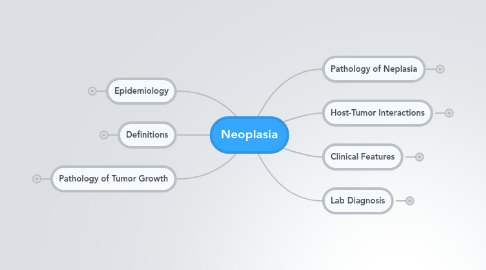
1. Epidemiology
1.1. MCC Cancer Deaths
1.1.1. Lung
1.1.2. Breast
1.1.3. Prostate
1.1.4. Colo-recal
1.2. MCC Cancer in Men
1.2.1. lung
1.2.2. prostate
1.2.3. colo-rectal
1.2.4. hematopoietic
1.2.5. urinary tract
1.2.6. pancreas
1.3. MCC Cancer in women
1.3.1. lung
1.3.2. breast
1.3.3. colo-rectal
1.3.4. hematpoietic
1.3.5. ovary
1.3.6. pancreas
1.4. MCC Cancer in Children
1.4.1. Leukemia
1.4.2. Lymphoma
1.4.3. Brain
1.4.4. Colo-rectal
1.4.5. Bone & Soft Tissue
1.4.6. Endocrine
1.5. Change in Incidence/Death
1.5.1. decline for GI
1.5.1.1. better food preservation
1.5.1.2. change in dietary habits
1.5.2. decline for breast, uterine, cervical
1.5.2.1. improved screening
1.5.2.2. early detection
1.5.3. lung cancer
1.5.3.1. reduce smoking prevalence
1.5.3.2. reduce environmental smoke exposure
1.5.4. inc incidence among ethinic groups
1.5.4.1. multifactorial
1.5.4.2. socioeconomic
1.5.4.3. environmental
1.5.4.4. genetic
1.6. Factors for Cancer incidence
1.6.1. hereditary/genetic predisposition
1.6.1.1. 5-10% of all cencer
1.6.1.2. autosomal dominant
1.6.1.2.1. single mutant gene
1.6.1.2.2. point mutation in single allele
1.6.1.2.3. mutation in second allele
1.6.1.2.4. tumors arise in specific sites
1.6.1.2.5. specific marker phenotype
1.6.1.3. autosomal recessive
1.6.1.3.1. defective DNA repair
1.6.1.3.2. very rare
1.6.1.3.3. xeroderma pigmentosum
1.6.1.3.4. ataxia-telangiectasia
1.6.1.3.5. bloom syndrome
1.6.1.3.6. fanconi anemia
1.6.1.4. familial cancer
1.6.1.4.1. occur at higher frequency in famlies
1.6.1.4.2. pattern not clearly defined
1.6.1.4.3. siblings - 2-3x great risk
1.6.1.4.4. no marker phenotpe
1.6.1.4.5. early onset < 40
1.6.1.4.6. multiple/bilateral tumors
1.6.1.4.7. fam hx of cx in 2 or more 1/2 degree
1.6.2. age
1.6.3. social habits
1.6.4. environmental/occupational
1.6.5. pre-existing disease states
1.6.6. chemo-radiation therapy
2. Definitions
2.1. Hyperplasia
2.1.1. inc in cell @
2.2. Hypertrophy
2.2.1. cell enlargement without division
2.3. metaplasia
2.3.1. irreversible change of one mature type to another
2.4. Neoplasia
2.4.1. new abnormal growth (tumor)
2.4.1.1. proliferative
2.4.1.2. uncoordinated
2.4.1.3. autonomous
2.4.1.4. genetically unstable
2.4.1.5. composed of
2.4.1.5.1. proliferating neoplastic parenchyma
2.4.1.5.2. fibro-vascular supporting stroma
2.5. polyp
2.5.1. abnormal growth
2.5.2. from mucosal surface
2.5.3. attached by stalk
2.6. desmoplasia
2.6.1. dense fibrous stroma
2.6.2. seen w malignant tumors
2.6.3. invasion into ECM
2.6.4. altered stroma
2.6.4.1. activated fibroblasts
2.6.4.2. cytokines
2.6.4.3. growth factors
2.6.5. inc collagen deposition
2.6.6. histology
2.6.6.1. dense
2.6.6.2. pink
2.6.6.3. fibrotic
2.6.6.4. desmoplasia
2.7. Benign tumors
2.7.1. Examples
2.7.1.1. -oma
2.7.1.2. epithelial
2.7.1.2.1. cell of origin
2.7.1.2.2. microscopic pattern
2.7.1.2.3. macroscopic architecture
2.7.1.2.4. Papilloma
2.7.1.2.5. Hamartoma
2.7.1.2.6. Adenoma
2.7.2. slow growing
2.7.3. well differentiated
2.7.4. localized (encapsulated margins)
2.7.5. Tx: surgical excision
2.7.6. may have lethal complications
2.7.6.1. meningioma - hydrocephalus
2.7.6.2. gastric leioma - hemorrhage
2.7.6.3. insulin-producing pancreatic tumor
2.7.6.4. insulinoma - hypoglycemia
2.7.6.5. atrial myxoma - cardiac obstruction
2.7.7. exceptions
2.7.7.1. capillary hemangiomas
2.7.7.1.1. invasive margins
2.7.7.2. pituitary adenomas
2.7.7.2.1. visual disturbances
2.8. Malignant tumors
2.8.1. Nomenclature
2.8.1.1. cells of origin
2.8.1.1.1. Carcinoma
2.8.2. capable of invasion
2.8.3. capable of metastasis
2.8.4. rapid growing
2.8.5. moderate/poorly differentiated
2.8.6. desmoplastic stroma
2.8.6.1. example
2.8.7. Microscopic Characteristics
2.8.7.1. cellular & architectural pleomorphis
2.8.7.1.1. loss of organized growth
2.8.7.2. mitotic/apoptotic activity
2.8.7.2.1. abnormal mitotic activity
2.8.7.3. tumor necrosis
2.8.7.3.1. example
2.8.7.4. stromal & lympho-vascular invasion
3. Pathology of Neplasia
3.1. Differentiation
3.1.1. morphologic/physiologic characteristics
3.1.2. structure & function concordant
3.1.3. benign tumors
3.1.3.1. well differntiatied
3.1.4. malignant tumors
3.1.4.1. range of differntiation
3.1.4.2. increased proliferative act
3.1.4.3. variable loss of functional properties
3.1.4.4. anaplasia
3.1.4.4.1. little to no evidence of differentiation
3.2. Dysplasia
3.2.1. disordered growth
3.2.2. epithelium or mucosa
3.2.3. genetic changes
3.2.4. precursor to cancer
3.2.5. result in loss of
3.2.5.1. maturation
3.2.5.2. polarity
3.2.5.3. architectural orientation
3.2.6. pleomorphism
3.2.6.1. abnormalities in shape and size
3.2.6.2. example
3.2.7. increased mitotic activity
3.2.8. carcinoma in situ
3.2.8.1. premalignant/non-invasive state
3.2.8.2. full epithelial thickness of dysplasia
3.2.8.3. no invasion of the basement membrane
3.2.9. invasive carcinoma
3.2.9.1. dysplastic cells breach basement membrane
3.2.10. example
3.2.11. example
3.3. Tumor Grade
3.3.1. level of morphologic differntation
3.3.1.1. Example: Colon
3.3.1.1.1. well
3.3.1.1.2. moderately
3.3.1.1.3. poorly
3.3.2. categories of evaluation
3.3.2.1. size & shape
3.3.2.2. nuclear:cytoplasmic ratio
3.3.2.2.1. large nucleoli
3.3.2.3. nuclear and nucleolar characteristics
3.3.2.3.1. hyperchromasia
3.3.2.4. cytoplasmic differentation
3.3.2.5. mitotic activity
3.3.2.6. tissue organization
3.3.2.7. necrosis
3.3.2.8. lymphovascular invasion
3.4. Tumor Stage
3.4.1. distribution and extent of cancer @ time of diagnosis
3.4.2. tumor characteristics
3.4.2.1. tumor size
3.4.2.2. location
3.4.2.3. depth & extent of local invasion
3.4.3. nodal involvement
3.4.4. Metastases
3.4.5. aids in selection of therapy
3.5. Carcinogenesis
3.5.1. Agents
3.5.1.1. chemical carcinogens
3.5.1.1.1. examples
3.5.1.2. radiant energy
3.5.1.2.1. inc cutaneous cancer
3.5.1.2.2. UVB (280-320 nm)
3.5.1.2.3. UVC (200-280 nm)
3.5.1.2.4. ionizing radiation
3.5.1.3. oncogenic viruses & microbes
3.5.1.3.1. Human Papilloma Virus
3.5.1.3.2. Epstein Barr Virus
3.5.1.3.3. Hep B
3.5.1.3.4. Human T-Cell Leukemia (RNA) virus type 1
3.5.1.3.5. H. pylori
3.5.1.4. in vivo
3.5.1.4.1. mutagens in virtro
3.5.2. initiation
3.5.2.1. irreversible
3.5.2.2. injures DNA
3.5.2.2.1. cellular mutation
3.5.2.3. procarcinogens
3.5.2.3.1. indirectly damage DNA following metabolic activation
3.5.3. promotion
3.5.3.1. promoters
3.5.3.1.1. stimulate division of initiated cell
3.5.3.1.2. exogenous
3.5.3.1.3. endogenous
3.5.3.2. reversible
3.5.3.3. non-tumorgenic
3.5.3.4. proliferative
3.5.3.5. induces tumor formation
3.5.3.6. time dependent
3.5.4. progression
3.5.4.1. autonomous growth
3.5.4.2. genomic instability
3.5.4.3. tumor heterogeneity
3.5.5. cancer
3.5.5.1. invasion
3.5.5.2. metastasis
3.5.6. summary
3.5.6.1. carcinogenesis
4. Pathology of Tumor Growth
4.1. Rate
4.1.1. morphologic grade
4.1.2. mitotic activity
4.1.3. nature of neoplasm
4.1.4. supportive stroma & neovascularization
4.1.5. GF & hormone depenence
4.1.6. interventional treatment
4.2. Phases
4.2.1. Monoclonal proliferations
4.2.1.1. growth & genetic instability
4.2.1.2. Progression
4.2.1.2.1. subpopulations
4.2.2. clonal growth and malignant change
4.2.3. local growth and angiogensis
4.2.4. regional invasion
4.2.5. metastatic spread
4.2.5.1. marks a tumor as malignant
4.2.5.2. 20-30% new dx have mestasis
4.2.6. growth fraction
4.2.6.1. proliferating component
4.2.6.2. minority of tumor cells
4.2.6.3. most susceptible to cell-cycle based chemotherapy
4.2.6.4. < 20% of tumor
4.3. Infiltrative Growth Margin
4.3.1. local destructive invasion
4.3.2. direct seeding
4.3.2.1. body cavities
4.3.2.1.1. peritoneal
4.3.2.1.2. pericardial
4.3.2.1.3. pleural
4.3.2.2. example
4.3.3. lymphatic spread
4.3.3.1. MC for carcinoma
4.3.3.2. example
4.3.4. hematogenous spread
4.3.4.1. MC for sarcoma
4.3.4.2. example
4.3.5. clinical presentation
4.3.5.1. most @ Site of origin
4.3.5.2. 20% metastatic distant site
4.4. Development
4.4.1. Growth factors
4.4.1.1. TGFbeta
4.4.1.2. bFGF
4.4.1.3. PDGF
4.4.1.4. VEGF
4.4.2. Host factors
4.4.2.1. angiogenesis
4.4.3. Proteinases
4.4.3.1. digest basement membrane
4.4.3.2. enable cell infiltration
4.4.3.3. digest stromal matrix
4.5. Invasion
4.5.1. Invasion of Extracellular Matrix
4.5.1.1. Loss of Adhesion molecules
4.5.1.2. down regulation of cadherins
4.5.1.3. attachment to matrix via integrins
4.5.1.4. degradation of ECM
4.5.1.5. detachment from solid mass
4.5.1.6. move into circulation
4.5.2. Vascular Dissemination/Horning of Tumor Cells
4.5.2.1. Intravasation
4.5.2.1.1. intrxn w host immune system
4.5.2.2. Dissemination
4.5.2.2.1. bind to blood cells
4.5.2.2.2. pass basement membrane
4.5.2.3. Extravasation
4.5.2.3.1. Micrometastasis
4.5.2.3.2. Colonization
4.6. Metastatic sites
4.6.1. determined by
4.6.1.1. anatomic
4.6.1.2. vascular
4.6.1.3. host-tumor paracrine effects
4.6.1.4. tropism certain organs
4.6.1.4.1. adhesion molecules for vascular bed
4.6.2. Brain
4.6.2.1. lung
4.6.2.2. breast
4.6.2.3. kidney
4.6.2.4. GI
4.6.2.5. melanoma
4.6.3. Liver
4.6.3.1. GI
4.6.3.2. pancreato-biliary
4.6.3.3. breast
4.6.3.4. lung
4.6.4. bone
4.6.4.1. prostate
4.6.4.2. breast
4.6.4.3. lung
4.6.4.4. kidney
4.6.4.5. thyroid
4.6.4.6. testes
5. Host-Tumor Interactions
5.1. Tumor Antigens
5.1.1. Tumor specific (tumor cells only)
5.1.2. Tumor associated (tumor/normal cells)
5.1.3. classification
5.1.3.1. molecular structure
5.1.3.2. source
5.1.4. characteristics
5.1.4.1. normal cellular proteins expressed abnormally
5.1.4.2. mutated genes
5.1.4.3. oncogenic viruses
5.1.4.4. high levels on cancer cells
5.1.4.5. altered glycolipids/glycoproteins
5.1.4.6. present on cells of origin
5.2. Immunity
5.2.1. Cell mediated immunity
5.2.1.1. dominant antitumor mechanism
5.2.1.2. CD8+ cytotoxic T cell
5.2.1.3. recognized MHCI
5.2.1.4. NK cells
5.2.1.5. Macrophages
5.2.1.6. Antibodies
5.2.2. Immune Surveillance
5.2.2.1. inc in immunocompromised host
5.2.2.2. can escape immune surveillance
5.2.2.2.1. Ag-neg variants
5.2.2.2.2. red MHC
5.2.2.2.3. lack co-stim
5.2.2.2.4. immunoosuppression
5.2.2.2.5. Ag masking
5.2.2.2.6. CD8+ apoptosis
6. Clinical Features
6.1. clinical presentation
6.1.1. asymptomatic mass
6.1.2. pain/tenderness
6.1.3. non-healing lesino
6.1.4. persistent cough
6.1.5. abnormal bleeding
6.1.6. peforation/obstruction
6.1.7. non-specific weakness
6.1.8. fatigue
6.2. mechanisms
6.2.1. effect of compression/obstruction
6.2.2. secretory/physiological effects
6.2.2.1. pituitary adenoma
6.2.2.1.1. compress optic nerve
6.2.2.1.2. visual disturbances
6.2.2.1.3. hormone overexpression
6.2.3. bleeding due to ulceration
6.2.4. vascular injury
6.2.5. secondary infection
6.2.6. cachexia
6.2.6.1. weakness
6.2.6.2. anorexia
6.2.6.3. weight loss
6.2.6.4. anemia
6.2.6.5. cause of 30% mortalitiy
6.2.6.6. result of TNF from macrophages
6.3. endocrine/neuroendocrine tumors
6.3.1. absence of function of hormone
6.3.2. overproduction of hormone
6.3.3. suppression of hormonal activity
6.3.4. overstimulation of hormonal activity
6.4. Tumor markers
6.4.1. detection
6.4.1.1. tumor cells
6.4.1.2. serum
6.4.1.3. effusions
6.4.2. indicators of specific cancer
6.4.3. confirm diagnosis
6.4.4. assess tumor staging
6.4.5. monitor for response to therapy
6.4.6. ID recurrent disease
6.4.7. examples
6.4.7.1. PSA
6.4.7.1.1. prostatic cancer
6.4.7.2. beta-HCG
6.4.7.2.1. gestational trophoblastic disease
6.4.7.2.2. choriocarcinoma
6.4.7.2.3. gonadal cancer
6.4.7.3. alpha fetoprotein
6.4.7.3.1. testicullar cancer
6.4.7.3.2. hepatocellular cancer
6.4.7.4. CEA
6.4.7.4.1. GI
6.4.7.4.2. Pancreato-biliary
6.4.7.5. CA-125
6.4.7.5.1. oavarian
6.5. Paraneoplastic Syndroms
6.5.1. seconary to tumor progression
6.5.2. hormones or factors
6.5.3. earliest manifestation of occule malignancy
6.5.4. 10% of patients
6.5.5. presentation
6.5.5.1. endocrinopathy
6.5.5.2. hypercalcemia
6.5.5.3. neuromuscular deficiency
6.5.5.4. dermatologic conditions
6.5.5.5. bone changes
6.5.5.6. hematologic abnormalities
6.5.6. Tx: address primary deficiency
6.5.7. examples
6.5.7.1. Cushing syndrome
6.5.7.1.1. ACTH
6.5.7.1.2. small cell carcinoma of lung
6.5.7.1.3. pancreatic carcinoma
6.5.7.1.4. neural tumors
6.5.7.2. Syndrome of Inappropriate Antidiuretic Hormone Secretion
6.5.7.2.1. ADH/atrial natriuretic hormone
6.5.7.2.2. small carcinoma of lung
6.5.7.2.3. intracranial neoplasms
6.5.7.3. Hypercalcemia
6.5.7.3.1. Parathyroid hormone related protein
6.5.7.3.2. Squamous cell carcinoma of lung
6.5.7.3.3. breast
6.5.7.3.4. renal
6.5.7.3.5. adutl T-cell leukemia/lymphoma
6.5.7.3.6. ovarian carcinoma
6.5.7.4. carcinoid syndrome
6.5.7.4.1. Serotonin
6.5.7.4.2. Bradykinin
6.5.7.4.3. carcinoid tumor
6.5.7.4.4. gastric carcinoma
6.5.7.4.5. pancreatic carcinoma
7. Lab Diagnosis
7.1. Clinical history
7.2. physical examination
7.3. radiologic imaging
7.4. lab tests
7.5. pathologic exam
7.5.1. surgical methods
7.5.1.1. Biopsy
7.5.1.2. excision
7.5.1.3. resection
7.5.1.4. rapid frozen section
7.5.1.4.1. at time of surgery
7.5.2. cytologic methods
7.5.2.1. Pap smears
7.5.2.2. Fine Needle Aspiration
7.5.3. ultrastructural methods
7.5.3.1. electron microscopy
7.5.4. molecular methods
7.5.4.1. immunohistochemistry
7.5.4.1.1. technique
7.5.4.1.2. function
7.5.4.2. flow cytometry
7.5.4.3. FISH
7.5.4.4. PCR
7.5.4.5. based on genetic makeup/expression
7.5.4.6. can alter treatment
7.5.4.7. more accurate testing/prognosis

