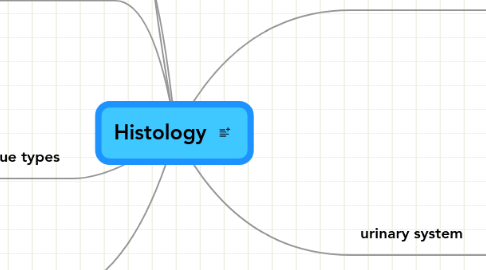
1. vascular system
1.1. veins
1.1.1. general characteristics
1.1.2. venules
1.2. arteries
1.2.1. 3 basic layers
1.2.1.1. tunica intima
1.2.1.2. tunica media
1.2.1.3. tunica adventitia
1.2.2. arterioles
1.3. capillaries
1.3.1. general characteristics
1.3.2. 3 basic types
1.3.2.1. Type I - continuous
1.3.2.2. Type II- fenestrated
1.3.2.3. Type III- discontinuous
1.4. lymphatic system
1.4.1. primary functions
1.4.2. lympathic vessels
1.4.3. lymphoid tissue
1.4.3.1. lymphocytes
1.4.3.2. diffuse
1.4.3.3. nodules
1.4.4. lymphatic organs
1.4.4.1. lymph node
1.4.4.2. spleen
1.4.4.3. thymus
1.4.4.4. tonsils
2. digestive system
2.1. accessory glands
2.1.1. salivary glands
2.1.1.1. parotid
2.1.1.2. submandibular
2.1.1.3. sublingual
2.1.2. pancreas
2.1.3. liver
3. four tissue types
3.1. epithelial tissue
3.1.1. membrane epithelia p. 7
3.1.1.1. simple squamous
3.1.1.1.1. barrier that is easily penetrated
3.1.1.1.2. endothelia in blood vessels
3.1.1.1.3. mesothelia in visceral organs
3.1.1.1.4. lung alveoli
3.1.1.2. simple cuboidal
3.1.1.2.1. slightly more substantial barrier than simple squamous but still allowing significant movement across the cells
3.1.1.2.2. kidney tubules
3.1.1.2.3. exocrine gland ducts
3.1.1.3. stratified squamous
3.1.1.3.1. thicker layer that resists physical abrasion
3.1.1.3.2. keratinized: epidermis
3.1.1.3.3. non-keratinized: oral cavity, esophagus, anal canal
3.1.1.4. pseudostratified
3.1.1.4.1. mostly columnar cells, some goblet cells
3.1.1.4.2. bronchi, trachea
3.1.1.5. stratified cuboidal (rare)
3.1.1.5.1. exocrine glands, fetal epidermis
3.1.1.6. stratified columnar (rare)
3.1.1.6.1. male prostatic urethra
3.1.1.7. transitional
3.1.1.7.1. mostly stratified squamous looking with cuboidal surface layer that stretches upon pressure
3.1.1.7.2. urinary bladder, ureter, parts of kidney
3.1.2. glandular epithelia
3.2. nervous tissue
3.2.1. central nervous system
3.2.1.1. general concepts
3.2.1.1.1. glial cells
3.2.1.1.2. gray/white matter
3.2.1.1.3. meninges
3.2.1.2. divisions
3.2.1.2.1. spinal cord
3.2.1.2.2. cerebrum
3.2.1.2.3. cerebellum
3.2.2. peripheral nervous system
3.2.2.1. myelination by schwann cells
3.2.2.2. ganglia
3.2.2.2.1. sensory (afferent)
3.2.2.2.2. autonomic (efferent)
3.2.2.3. nerve structure
3.2.2.3.1. axons
3.2.2.3.2. fascicles
3.2.2.3.3. perineurium
3.2.2.3.4. 2nd degree bundles
3.2.2.3.5. endoneurium
3.2.3. general characteristics
3.2.3.1. neurons
3.2.3.1.1. axons
3.2.3.1.2. Nissl bodies - rER
3.2.3.1.3. perikaryon - cell body
3.2.3.1.4. dendrites
3.2.3.2. synapses
3.2.3.2.1. axo-dendritic
3.2.3.2.2. axo-axonic
3.2.3.2.3. axo-somatic
3.2.3.2.4. myoneural
3.3. connective tissue
3.3.1. composition
3.3.1.1. matrix
3.3.1.1.1. amorphous ground substance
3.3.1.1.2. GAG's
3.3.1.2. cells
3.3.1.2.1. adipose
3.3.1.2.2. plasma
3.3.1.2.3. fibroblasts
3.3.1.2.4. blood
3.3.1.2.5. bones
3.3.1.2.6. mast
3.3.1.2.7. macrophages
3.3.1.2.8. chondroblasts
3.3.2. classification
3.3.2.1. general CT
3.3.2.1.1. dense
3.3.2.1.2. non dense
3.3.2.2. special CT
3.3.2.2.1. cartilage
3.3.2.2.2. bone
3.3.2.2.3. blood
3.4. muscles
3.4.1. classification
3.4.1.1. skeletal
3.4.1.2. smooth
3.4.1.3. cardiac
3.4.2. structural hierarchy
3.4.2.1. muscle fasciculus
3.4.2.2. myofibers/myocytes
3.4.2.3. myofibrils
3.4.2.4. myofilaments
3.4.2.4.1. actin filament (thin)
3.4.2.4.2. myosin filament (thick)
3.4.2.5. sarcomere
4. reproductive system
4.1. female
4.1.1. ovary
4.1.1.1. oogenesis
4.2. male
5. My Geistesblitzes
5.1. immunology
6. respiratory system
6.1. tracheea
6.2. bronchus
6.3. bronchioles
7. urinary system
7.1. kidneys
7.1.1. macro structures
7.1.1.1. cortex
7.1.1.2. medulla
7.1.1.2.1. medullary pyramids
7.1.1.2.2. area cribrosa
7.1.1.3. renal sinus
7.1.1.3.1. pelvis
7.1.1.3.2. calyces
7.1.2. blood supply
7.1.2.1. renal artery
7.1.2.2. interlobar artery
7.1.2.3. arcuate artery
7.1.2.4. interlobular artery
7.1.2.5. intralobular artery
7.1.2.6. afferent arteriole
7.1.2.7. glomerulus
7.1.2.8. efferent arteriole
7.1.3. micro structures
7.1.3.1. nephrons
7.1.3.1.1. types
7.1.3.1.2. components
