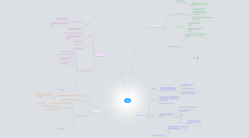
1. Nervuous Tissue
1.1. Function
1.1.1. Recieves stimuli and conducts nerve impulses
1.1.1.1. Nervous system has three functions: sensory input, integration of data, and motor outout
1.2. Found
1.2.1. In the brain and spinal cord (nervous system)
1.3. Appearance/ Composition
1.3.1. Contains neurons: specialized nervous cells
1.3.1.1. Nervous System
1.3.2. Contain Neuroglia
1.3.2.1. Nine times more in number than neurons
1.3.2.2. Support and nourish neutrons
1.3.3. Picture
2. Epithelial Tissue a.k.a Epithelium
2.1. Function
2.1.1. Mainly for protection
2.1.1.1. External: from injury, drying out, pathogen invasion
2.1.1.2. Internal: secrets mucus along digestive tract and sweeps up impurities from the lungs
2.1.2. Also for secretion, absorption, excretion, and filtration
2.2. Found
2.2.1. Covers body surfaces and lines boy cavities
2.2.2. In the lining of the lungs, blood vessels, kidney tubules, digestive tracts, and oviducts
2.3. Types
2.3.1. Squamous epithelium
2.3.2. Cubodial epithelium
2.3.3. Columnar epithelium
2.4. Appearance/ Composition
2.4.1. Occur in more than one layer
2.4.2. Named according to the shape of the cell
2.4.3. Cells lining a body cavity can be ciliated and/or grandular
2.4.4. Tightly packed cells that form a continous layer
2.4.5. Picture
3. Muscular (Contractile) Tissue
3.1. Function
3.1.1. Moves the body and its parts
3.2. Types
3.2.1. Skeletal Muscle a.k.a. voluntary muscle
3.2.1.1. Found: Attached by tendons to the bones of the skeleton (when it contracts, body moves)
3.2.1.2. Apperance: Cylindrical an long with the nuclei located at the periphery of the cell
3.2.2. Smooth (Visceral) Muscle
3.2.2.1. Unlike skeletal muscle, muscle is involuntary (not under voluntary control), is slower to contract but stays contracted for a longer period of time
3.2.2.2. Found: In the walls of the intestine, stomach, and other internal organs. As well as, found in blood vessels
3.2.3. Cardiac Muscle
3.2.3.1. Found: only in the walls of the heart (contraction controls heartbeat)
3.2.3.2. Appearance: branched cells, have single (central) nucleus, has striations
3.3. Appearance/ Composition
3.3.1. Composed of muscle fibers (cells) which contain actin filaments and myosin filaments (cause movement)
3.3.2. Picture
4. Connective Tissue
4.1. Function
4.1.1. Binds organs together, provides support and protection, fills spaces, produces blood cells, and stores fat
4.1.1.1. i.e. Protection for epithelium and internal organs
4.1.1.2. Can form protective covering
4.1.1.3. Connect muscles to bones, and bones to other bones and joints
4.2. Found
4.2.1. In the lungs, arteries, urinary bladder, internal organs, tendons, ligaments, beneath the skin, around the kidneys, on the surface of the heart, lymph nodes
4.2.2. Are cartilage, bones, and blood
4.3. Types
4.3.1. Differ according to thype of matrix and the abundance of fibers in the matrix
4.3.1.1. Loose Fibrous and Dense Fibrous Tissues
4.3.1.2. Adipose Tissue and Reticular Connective Tissue
4.3.1.3. Cartilage
4.3.1.4. Bone
4.3.1.5. Blood
4.3.1.5.1. Not all people classify this as a connective tissue (vascular tissue instead)
4.4. Appearance/ Composition
4.4.1. Are widely seperated by a matrix (made up of a noncellular material that can vary from being solid to semifluid to fluid
4.4.2. Connective Tissue
