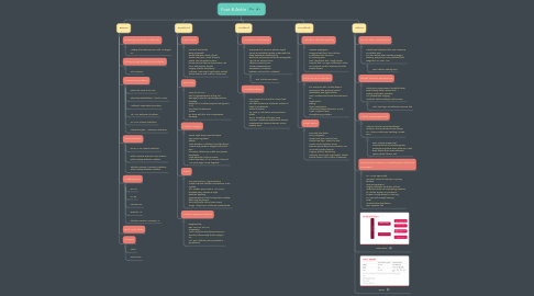
1. Basics
1.1. Resting position subtalar:
1.1.1. Midway b/w extreme ROM with 10 degree PF
1.2. Close pack position subtalar:
1.2.1. Full inversion
1.3. Capsular pattern
1.3.1. Talocrural: Loss of PF> DF
1.3.2. Talocalcaneal(subtalar): > limit in varus
1.3.3. Midtarsal: Supination>pronation
1.3.4. 1st MTP: Extension limitation
1.3.5. 2-5 MTP: Flexion restriction
1.3.6. Interphalangeal : extension restriction
1.4. Goniometry
1.4.1. DF, PF AXIS: Lateral malleolus
1.4.2. ankle Invesion, eversion Axis: anterior ankle midway between malleoli
1.4.3. Subtalar eversion, inversion: posterior ankle midway between malleoli
1.5. Ankle ROM
1.5.1. DF: 20
1.5.2. PF: 50
1.5.3. Inversion 35
1.5.4. Eversion 15
1.5.5. Subtarar eversion, inversion: 5
1.6. Roll and slide
1.7. nerves
1.7.1. Tibial
1.7.2. Sural nerve
2. Forefoot
2.1. Heel Spur
2.1.1. Pain with heel strike Bony outgrowth Dorsal heel spur: back of heel Plantar heel spur: under the sole Cause: can be systemic issue; biomechanical-abnormal pronation; can occur with plantar fasciitis Surgery: plantar fasciotomy Orthosis: Heel insert, night splint, stretch Plantar flexors, soft cushion, ultrasound
2.2. Turf Toe
2.2.1. Injury of 1st MTP Due to hyperextension of big toe Damage to plantar capsuloligamentous complex Player fall on another player’s heel (grade I, II, III) Pain distal to sesamoid Tender Rx- shoe with firm sole, compressive bandage
2.3. Hallux valgus
2.3.1. Cause: tight shoes, pointed shoes Pain onset is gradual Effects: -Gait deviation- midstance and late stance -Lateral and posterior weight shift due to pain -Pronation deformity, unable to supinate, pes planus Weak abductor hallucis muscle Lateral deviation of 1st MTP joint (Normal MTP joint angle =8-20 degrees)
2.4. Gout
2.4.1. Uric acid level inc., hyperuricemia Crystal induced arthritis-monosodium urate crystals S/S- Sudden pain onset in 1st MTP jt., localized pain, intense at night Redness, swelling Hypersensitivity, chills, tachycardia, malaise, fever may be present Skin-red/purple, shiny, tense, warm Drugs: Allopurinol, colchicine, probenazide
2.5. Hallux rigidus/limitus
2.5.1. Degenerative Dec. ROM at 1st MTP Progressive MOI: Faulty function/biomechanics or structural abnormality (hallux valgus) OA S/S- Pain, stiffness, pain provoked in Dorsiflexion.
3. Midfoot
3.1. Morton’s Neuroma
3.1.1. Entrapment of common plantar digital nerves & repetitive traction underneath the deep transverse metatarsal lig. Epineural and perineural fibrous overgrowth Age 45-50, women>men Insidious onset of pain Plantar webspace pain Paresthesia, numbness between 3rd and 4th Metatarsal
3.1.1.1. Test: Plantar percussion
3.2. Metatarsalgia
3.2.1. Pain, proximal to toes b/w 1st and 2nd MTP joint. Pain and tenderness of plantar surface of head of metatarsals Callus formation Inc. pain in mid stance and propulsion phase Rx-inc. Flexibility of triceps surae Orthosis- metatarsal pad(internal device), metatarsal bar (external device), rocker bottom shoe.
4. Hindfoot
4.1. Achilles Tendinopathy
4.1.1. Traction apophysitis Overpronated foot, low nutrition In athletes more common s/s morning pain Test: Thompson test , Royal london hospital test, Arc sign, Hoffa test, Matles Rx- Eccentric control exercises of ankle Plantar flexors
4.2. PLANTAR FASCITIS
4.2.1. s/s: Heel pain with 1st few steps in morning or after prolong limited dorsiflexion and tight achilles. Test: Windlass test-(Great toe extension) Rx Night Splint Taping Shoe modification Ultrasound (dexamethasone, 3MHz) UCBL, Scaphoid pad Strengthening inverters
4.3. Heel Spur
4.3.1. Pain with heel strike Bony outgrowth Dorsal heel spur: back of heel Plantar heel spur: under the sole Cause: can be systemic issue; biomechanical-abnormal pronation; can occur with plantar fasciitis Surgery: plantar fasciotomy Orthosis: Heel insert, night splint, stretch Plantar flexors, soft cushion, ultrasound
5. Other
5.1. Sinus Tarsi Syndrome
5.1.1. Tube/tunnel between talus and calcaneus or subtalar joint s/s: pain worse when standing, weight bearing, pes planus, over pronated foot Diagnosis: CT Scan, MRI
5.1.1.1. Test: subtalar stability test
5.2. Tarsal tunnel syndrome
5.2.1. Tibial nerve compression (medially) when pass through flexor retinaculum. History of Stressful activities C/C paresthesia, tingling Abductor hallucis atrophy can be seen
5.2.1.1. Test: Tinel sign, Dorsiflexion-eversion test
5.3. Ankle Impingement
5.3.1. Anterior: due to excess dorsiflexion Posterior: due to excess plantar flexion c/c : chronic ankle pain, swelling, limited ROM
5.3.1.1. Test: Anterior drawer test Silverskiold test of isolated gastroc contracture-positive when ankle DF> with knee flexion then Knee extension Hyper plantar flexion Test
5.4. Calcaneal Traction Apophysitis (Sever’s Disease)
5.4.1. In 7-15 yrs age mostly Common cause of heel pain in young athletes Overuse syndrome Osgood schlatter syndrome of foot Osteochondritis via overloading insertion of Achilles tendon on calcaneus s/s pain usually absent in morning Inc. pain with weight bearing Limp Limited ankle dorsiflexion Test: Squeeze test

