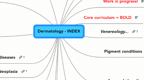
1. Eczemas
1.1. TYPES
1.1.1. Contact eczema
1.1.1.1. Allergic
1.1.1.1.1. ALLERGENS
1.1.1.1.2. PATHOGENESIS
1.1.1.1.3. TREATMENT
1.1.1.1.4. PICTURES
1.1.1.2. Nonallergic
1.1.1.2.1. PROGRESS
1.1.1.2.2. ETIOLOGY
1.1.2. Atopical eczema
1.1.2.1. PICTURE
1.1.2.2. DIAGNOSTICS
1.1.2.2.1. Itching skin disease + 3 of:
1.1.2.2.2. INVESTIGATION
1.1.2.2.3. Some clinical signs
1.1.2.2.4. Toddlers have...
1.1.2.2.5. Adults have...
1.1.2.3. ETIOLOGY
1.1.2.3.1. Altered Tcell function
1.1.2.3.2. Excess tendency of IgE production
1.1.2.3.3. Psychiatric factors
1.1.2.3.4. Scratching
1.1.2.4. COMPLICATIONS
1.1.2.4.1. Contact allergy
1.1.2.4.2. Herpex simplex eczema
1.1.2.4.3. Molluscum contagiosum
1.1.2.4.4. Impetigo
1.1.2.5. TREATMENT
1.1.2.5.1. Creams
1.1.2.5.2. Break the "itch circle"!
1.1.2.5.3. Light treatment
1.1.3. Nummular eczema
1.1.3.1. New node
1.1.4. Seborroic eczema
1.1.4.1. ETIOLOGY
1.1.4.1.1. Malassezia furfur (?)
1.1.4.1.2. Inherited
1.1.5. Localized types
1.1.5.1. Neurodermatitis
1.1.5.2. Diaper eczema
1.1.5.2.1. Picture
1.1.5.3. Statis eczema
1.2. GENERAL
1.2.1. Eczema = Epidermotitis = Inflamed skin
1.2.2. All eczemas are dermatites, but...
1.2.2.1. ...not all dermatites are eczemas!
2. Infections
3. Granulomatous diseases
4. Neoplasia
5. Papulo-squamous diseases
6. Leg wounds
6.1. Arterial
6.2. Venous
6.3. Anaesthetic
6.4. Purpura
6.5. BASIC KNOWLEDGE ABOUT:
6.5.1. Decubital wounds
6.5.2. Vasculitis
6.5.3. swe "Storkbett"
6.5.4. Nevus flammeus
6.5.5. Infantile hemangiomas
6.5.6. Telangiektasier
6.5.6.1. "Spider nevus"
6.5.7. Raynaud-fenomen
6.5.8. ”Cherry” angioma
6.5.9. Pyogenic granulomas
6.5.10. Lymph edemas
7. Autoimmune- / Systemic- / Blister-dermatose- diseases
8. Psychiatric diseases
8.1. Trichotillomania
8.1.1. Urge to pull out hair
8.1.2. a.k.a. "Trich" or TTM
9. Accumulative diseases
9.1. Xanthomas
9.2. Amyloidosis
9.3. Myxedema
10. Venereology...
11. Work in progress!
12. Hair, nails, adnex organs
13. Pigment conditions
13.1. Dark skin
13.2. Pigmental abberations
13.2.1. Speckled nevus
13.2.1.1. Pic (forehead of woman)
14. Hereditary conditions
15. INTRODUCTION
15.1. Diagnostics
15.1.1. BASIS
15.1.1.1. Patient history
15.1.1.2. Clinical examination
15.1.1.3. Lab results
15.1.1.3.1. Blood
15.1.1.3.2. Urine
15.1.1.3.3. Microbiology
15.1.1.3.4. Histopathology
15.1.2. TERMINOLOGY OF SKIN CHANGES
15.1.2.1. TISSUE GAIN
15.1.2.1.1. Vesicle (small blister)
15.1.2.1.2. Urtica (medium bump)
15.1.2.1.3. Papule (small bump)
15.1.2.1.4. Pustule (blister of pus)
15.1.2.1.5. Nodule (firm bump)
15.1.2.1.6. Tumor (firm elevation)
15.1.2.1.7. Hypertrophia
15.1.2.1.8. Crust
15.1.2.1.9. Bulla (blister)
15.1.2.1.10. Lichenification
15.1.2.2. Useful atlas here!
15.1.2.3. TISSUE LOSS
15.1.2.3.1. Excoriation
15.1.2.3.2. Scale (epidermal fragment)
15.1.2.3.3. Cicatrix
15.1.2.3.4. Sclerosis
15.1.2.3.5. Fissure
15.1.2.3.6. Atrophia
15.1.2.3.7. Erosion
15.1.2.3.8. Ulcus (wound)
15.1.2.4. NO GAIN / LOSS
15.1.2.4.1. Erythema (redding)
15.1.2.4.2. Macula (spot)
15.2. Skin basics
15.2.1. Function
15.2.1.1. Protection
15.2.1.2. Sensation
15.2.1.3. Production
15.2.1.3.1. of Vitamin D
15.2.1.4. Signalling
15.2.1.4.1. Social
15.2.1.4.2. Sexual
15.2.1.5. Heat reduction
15.2.2. Layers
15.2.2.1. Epidermis (top)
15.2.2.1.1. Barrier to the outside world
15.2.2.1.2. From 0.05 mm (eyelids) to >1mm (heels)
15.2.2.2. Dermis
15.2.2.2.1. CONTAINS
15.2.2.3. Subcutis (bottom)
15.3. ABBREVIATIONS
15.3.1. Abbreviations = less clutter!
15.3.2. BM = Basement Membrane
15.3.3. ED = EpiDermis
15.3.4. SC = SubCutis / SubCutaneous
15.3.5. 2' = Secondary (e.g. " 2' infection")
16. Core curriculum = BOLD
17. Urticaria etc
17.1. Urticaria
17.2. Angioedema
17.3. Anafylaxia
17.4. Toxikodermia
17.5. BASIC KNOWLEDGE OF:
17.5.1. Erythema multiforme
17.5.1.1. Minor
17.5.1.2. Major (Steven-Johnson syndrome)
17.5.1.2.1. Pic (hand on 6yo Boy)
17.5.2. Erythema nodosum
17.5.3. Fysikalisk urtikaria
17.5.4. Contact urticaria

