1. Atrial Fibrillation
1.1. Assessment: The rhythm will be irregularly irregular. The heart rate is typically in the 110-140 range. Atrial fibrillation may be chronic and is often associated with underlying heart disease such as rheumatic heart disease, atherosclerotic heart disease or congestive heart failure.
1.1.1. Treatment: If the patient is stable, obtain a 12-lead ECG and establish IV access. Consider rate control with diltiazem or beta blockers. If the patient is unstable cardiovert the patient at 50j and administer 20mg of Cardizem slow IVP. If needed cardiovert again at 100j and administer another 20mg of Cardizem slow IVP.
2. Ventricular Tachycardia (with a pulse)
2.1. Assessment: The heart rate is 100-250. The patient may be experiencing myocardial ischemia, increased sympathetic tone, hypoxia, idiopathic causes, acid-base disturbances, or electrolyte imbalances.
2.1.1. Treatment: CPR if needed. Cardiovert the patient at 100j and administer 150mg of amiodarone by slow IVP. Continue CPR, cardiovert at 100j, and give another 150 mg of amiodarone (only give two doses.) Give EPI 1/10,000 1mg every 3-5 minutes.
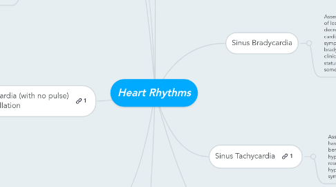
3. Ventricular Tachycardia (with no pulse) & Ventricular Fibrillation
3.1. Assessment: These are chaotic ventricular rhythms usually resulting from the presence of many reentry circuits within the ventricles. There are a wide variety of causes, most cases result from advanced coronary artery disease. There is no organized rate or rhythm.
3.1.1. Treatment: Start CPR. Cardiovert at 200j. Administer 300mg of amiodarone slow IVP. Administer EPI 1/10,000 1mg every 3-5 minutes. Start another round of CPR. Cardiovert at 200j. Administer another 150mg of amiodarone.
4. 1st degree Heart Block
4.1. Assessment: Rate will be 40-t0 BPM. An AV block can occur in the healthy heart, however, ischemia at the AV junction is the most common cause. The first degree block is usually no danger in itself.
4.1.1. Treatment: Provide oxygen, start an IV and consider atropine 0.5mg x3. TCP may be necessary. Start a dopamine drip if the BP is low after pacing.
5. 2nd degree Heart Block
5.1. Assessment: The rate is 40-50 BPM. There will be random QRS drops. Syncope and angina is common with a 2nd degree heart block.
5.1.1. Treatment: Provide oxygen, start an IV and consider atropine 0.5mg x3. TCP may be necessary. Start a dopamine drip if the BP is low after pacing.
6. Sinus Bradycardia
6.1. Assessment: The patient will have a heart rate of less than 60 with a regular rhythm. The decreases heart rate can cause decreased cardiac output, hypotension, angina, or CNS symptoms. In a healthy athlete, sinus bradycardia may be normal and have no clinical significance. Acute altered mental status and ongoing chest pain can also sometimes be seen.
6.1.1. Treatment: Treatment is usually unnecessary unless signs of poor perfusion are present. If there are signs of poor perfusion, prepare for transcutaneous pacing. Consider administering a 0.5mg bolus of atropine sulfate. Repeat every 3-5 minutes until you have obtained a satisfactory rate or have given 3.0 mg of the drug. If atropine fails, consider TCP or a dopamine or epinephrine infusion.
7. Sinus Tachycardia
7.1. Assessment: Heart rate is greater than 100 and has a regular rhythm. Sinus tachycardia is often benign. You may see hypovolemia, fever, or hypoxia in these patients. Sinus tachycardia can result from exercise, fever anxiety, hypovolemia, anemia, pump failure, increased sympathetic tone, hypoxia, or hyperthyroidism.
7.1.1. Treatment: Treatment is directed at the underlying cause. Hypovolemia, fever, hypoxia, or other causes should be corrected.
8. Atrial Flutter
8.1. Assessment: Atrial flutter may occur in normal hearts, but it is usually associated with organic disease. It rarely occurs as the direct result of an MI. Atrial dilation, which occurs with CHF, is a cause of atrial flutter. The atrial rate is 250-350 per minute.
8.1.1. Treatment: If the patient is stable, obtain a 12-lead ECG and establish IV access. Consider rate control with diltiazem or beta blockers. If the patient is unstable cardiovert the patient at 50j and administer 20mg of Cardizem slow IVP. If needed cardiovert again at 100j and administer another 20mg of Cardizem slow IVP.
9. 3rd degree Heart Block
9.1. Assessment: This is the absence of conduction between the atria and ventricles resulting from complete electrical block at or below the AV node. The 3rd degree heart block can result from MI, digitalis toxicity, or degeneration of the conductive system. These patients will be lethargic. The P-wave has no correlation to the QRS wave. The heart rate will be 20-30 BPM.
9.1.1. Treatment: Provide oxygen, start an IV and consider atropine 0.5mg x3. TCP may be necessary. Start a dopamine drip if the BP is low after pacing.
