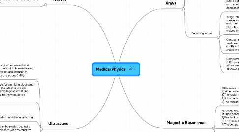
1. Tracers
1.1. Radioactive substances which are ingested or injected to measure metabolic functions or blood flow.
1.1.1. Beta Minus
1.1.1.1. Iodine-131-Has a half life of about 8.1 days,example of this is to measure the health of kidneys using a Geiger counter. Healthy kidneys will have a high count rate and then decrease , if there is a blockage the count rate will remain constant
1.1.2. Gamma
1.1.2.1. Technitium-99m-Half life of about 6 hours Used to monitor function of heart liver lungs kidneys etc. Leat ionising of radiation thus can pass through the patient with little loss of photons. And can be detected using a gamma camera
1.1.2.2. Gamma Camera 1)collimator which only allows gamma rays to be reached to the scintillator in one axis 2)Scintillator releases visible light photons onto a photo-cathode which releases an electron 3)Electron is accelerated to a dynode which releases another electron which are then accelerated towards futher dynodes producing an avalanche effect until it is a electrical pulse. 4)The computer then converts the signal to a display, each photomultiplier tube represents a pixel on the display
1.1.2.3. Positron emission tomography ( PET) , is used to measure brain functions 1) The patient is injected with a substance tagged with fluorine-18 (Usually glucose) 2)Fluorine 18 then decays a positron 3)Positron is then annihilated with an electron sending out two gamma rays in opposite directions 3)The pair of gamma rays are then detected by the PET scanner and using the time difference of detections, the position of the annihilation can be calculated. 4)Gradually a 3-D image of the brain is made.
2. Ultrasound
2.1. Ultrasound is any sound wave that is beyond the upperlimit of human hearing (20000 Hz).The ultrasound used in medical practice is around 2MHz
2.2. Ultrasound transducer is a device for emmiting ultrasound it consists of a piezoelectric crystal which gives out vibrations equal to the alternatic voltage across it and induces and e.m.f which is equal to the vibrations it recieves.
2.3. Acoustic impedance -density of material*the speed of sound in the material. -Radiographers are interested in the amount of US reflected at a boundary back to the receiver. It is dependent upon the acoustic impedance of the two materials at the boundary. -I/I0=(Z2-Z1/Z1=Z2)^2 -If the acoustic impendaces of the two materials are too different then the intensity recieved will be too great and wont be usefull to monitor deeper materials. An example of this is skin and air, a gel has to be used which is similar to the impedance of skin ,this is called impedance matching.
2.4. A scan- The e.m.f's induced can be plotted against a time graph to measure the thickness of a material,the time between two pulses is twice the distance traveled by the U.S.
2.5. B-Scan comprises of many A-Scans, the position and depth of reflecting surfaces is plotted to produce a 2-D cross sectional image of an organ.
2.6. Doppler effect -In the Doppler effect, the wavelength or frequency of a wave changes when there is a relative velocity between a source and detector -Can be used to measure blood flow as the transducer will receive u.s reflections from the iron rich blood. -The frequency of the reflected u.s will be different as it is moving away/closer to the transducer. -Blood flowing towards the transducer will have a higher frequency. -The difference in transmitted/received frequency corresponds to the velocity of blood flow.
3. Xrays
3.1. 10^-8 to 10^-13 m
3.2. Produced by accelerating electrons from a hot filament towards a tungsten anode about 1% of electrons are converted into xray photons
3.3. Three interaction mechanisms 1)Photoelectric effect <0.1 MeV ,electrons are absorbed by an electron which gains enough energy to be ejected from a target metal marterial 2)Compton scattering 0.5-5 MeV the photon looses some of its energy to an electon which is ejected from the metal 3)Pair production >1.02 MeV the photon entering an electric field of an atom produces a electron positron pair
3.4. Intensity of a beam of xrays is the power per unit area. I=Io*e^μx (μ is attenuation coefficient)
3.5. Detecting X-rays
3.5.1. Intensifier screens-the photons hit scintillating material such as phosphor which sends out visible light photons onto photographic film producing a brighter image, decreases exposure time by 100
3.5.2. Image intensifiers(Fluoroscopy)- Photon hits a phosphor screen, visible light photons hit a photo-cathode ejecting electrons towards an anode consisting of a small phosphor screen which gives out visible light (can be viewed on a monitor)
3.5.3. Contrast medium-Iodine/Barium is swallowed/injected it has a high attenuation coefficient (μ∝z^3)allowing us to outline shape of soft tissues such as intestines.
3.5.4. Computerized axial tomography (CAT)- Patient is placed in ring consisting of an x-ray tube and 720 x-ray detectors. The computer takes about 200 slices of images around the patient useful because- 1) Produces 3 dimensional image of the patient 2)Can distinguish between materials with similar attenuation co-efficients 3)Shows precise location ans shape of tumours.
