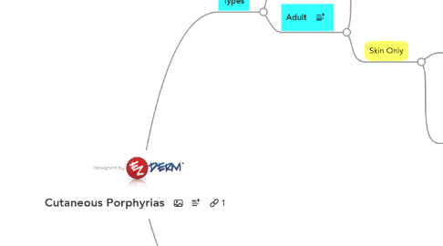
1. Types
1.1. Child
1.1.1. CEP
1.1.1.1. Mode of Inheritance
1.1.1.1.1. Autosomal Recessive
1.1.1.2. Enzyme Defect
1.1.1.2.1. Uroporphyrinogen III synthase
1.1.1.3. Clinical
1.1.1.3.1. Skin
1.1.1.3.2. Other
1.1.1.4. Labs
1.1.1.4.1. Uroporphyrin I predominates in urine
1.1.1.4.2. Erythrocyte porphyrins (EP) distinguish it from PCT (i.e., HEP)
1.1.1.4.3. Stool porphyrins used for monitoring
1.1.2. HEP
1.1.2.1. Mode of Inheritance
1.1.2.1.1. Autosomal Recessive (homozygous) form of Familial PCT
1.1.2.2. Enzyme Defect
1.1.2.2.1. Uroporphyrinogen decarboxylase
1.1.2.3. Clinical
1.1.2.3.1. Same as PCT
1.1.3. EPP
1.1.3.1. Mode of Inheritance
1.1.3.1.1. Autosomal Dominant
1.1.3.1.2. Autosomal Recessive (Heterozygous Mutation)
1.1.3.1.3. Chromosome Band: 18q21.3
1.1.3.2. Enzyme Defect
1.1.3.2.1. Ferrochelatase
1.1.3.3. Clinical
1.1.3.3.1. Painful phototoxicity due to Protoporphyrin IX
1.1.3.3.2. Caused by Visible Light
1.1.3.3.3. Petichiae, Purpura, Vesicles, Crusting
1.1.3.4. Labs
1.1.3.4.1. Can't PP with EPP
1.1.3.4.2. ZPP (Screening Test)
1.1.3.4.3. EP
1.1.4. Homozygous VP
1.1.4.1. Mutilating Photosensitivity
1.1.4.2. ZPP Elevated
1.2. Adult
1.2.1. Neurovisceral Symptoms
1.2.1.1. PBG
1.2.1.1.1. VP
1.2.1.1.2. HCP
1.2.2. Skin Only
1.2.2.1. PCT
1.2.2.1.1. Types
1.2.2.1.2. Precipitating Factors
1.2.2.1.3. Clinical Presentation
1.2.2.1.4. Associations
1.2.2.1.5. Treatment
1.2.2.1.6. Labs
1.2.2.2. Pseudo-PCT
1.2.2.2.1. Causes
1.2.2.2.2. Associations
1.2.2.2.3. 2 Clinical Patterns
1.2.2.2.4. Labs
2. Labs
2.1. Algorithmic Testing
2.1.1. Photosensitivity
2.1.1.1. Skin Only
2.1.1.1.1. Child
2.1.1.1.2. Adult
2.1.1.2. Neurovisceral + Skin
2.1.1.2.1. PBG
2.2. By Source
2.2.1. Blood
2.2.1.1. Erythrocyte porphyrins (EP)
2.2.1.1.1. CEP
2.2.1.1.2. EPP
2.2.1.2. ZPP (Screening Test)
2.2.1.2.1. EPP
2.2.1.2.2. HEP
2.2.1.3. Red-cell uroporphyrinogen decarboxylase activity
2.2.1.3.1. PCT
2.2.2. Urine
2.2.2.1. PBG
2.2.2.1.1. HCP
2.2.2.1.2. VP
2.2.2.2. Urine Porphyrins
2.2.2.2.1. Wood's Lamp gives Pink fluorescence
2.2.3. Feces
2.2.3.1. Stool Copro
2.2.3.1.1. HCP
2.2.3.2. Stool Proto
2.2.3.2.1. VP
2.3. By Disease
2.3.1. CEP
2.3.1.1. Uroporphyrin I predominates in urine
2.3.1.2. Erythrocyte porphyrins (EP) distinguish it from PCT (i.e., HEP)
2.3.1.3. Stool porphyrins used for monitoring
2.3.2. EPP
2.3.2.1. Can't PP with EPP
2.3.2.2. ZPP (Screening Test)
2.3.2.3. EP
2.3.3. Homozygous VP
2.3.3.1. ZPP Elevated
2.3.4. VP
2.3.4.1. PBG
2.3.4.2. Stool Protoporphyrinogen
2.3.4.3. Fluorescence Emission Peak (625-627 nm)
2.3.4.4. Urine (Copro > Uro)
2.3.5. HCP
2.3.5.1. Stool Coproporphyrinogen
2.3.5.2. Urine (Copro > Uro)
2.3.6. PCT
2.3.6.1. 24hr Urine Porphyrins
2.3.6.1.1. Wood's Lamp gives Pink fluorescence
2.3.6.2. Red-cell uroporphyrinogen decarboxylase activity
2.3.7. Pseudo-PCT
2.3.7.1. No porphyrin abnormalities
2.3.7.2. Exclude Connective Tissue Disease
