1. Risk factors
1.1. Other medical conditions
1.1.1. HIV infection
1.1.1.1. Number 1 risk factor
1.1.1.2. Since it weakens immune system, increases risk for TB
1.1.1.3. HIV + TB infection= HIV/TB coininfection
1.1.1.3.1. HIV medication + TB treatment is called antiretroviral therapy (ART)
1.1.1.4. TB disease may worsen HIV infection
1.1.1.4.1. Latent TB more likely to become a disease in those w/ HIV than those w/o HIV
1.1.2. Silicosis
1.1.2.1. Caused by exposure to silica dust
1.1.2.1.1. Prevalent in young people
1.1.2.1.2. Prevalent in low-income countries
1.1.3. Diabetes mellitus
1.1.3.1. Affects disease presentation and response
1.1.3.1.1. TB induces glucose intolerant and worsens glycaemic control
1.1.4. Organ transplants
1.1.4.1. Latent M tuberculosis found in transplants of brain, bone, kidney
1.1.5. Head or neck cancer
1.1.5.1. Weakened immune system
1.1.6. Specialized treatment for rheumatoid arthritis
1.1.6.1. Linked with treatment disturbances
1.1.7. Specialized treatment for Crohn's disease
1.1.7.1. Linked with treatment disturbances
1.2. Lifestyle/environment factors
1.2.1. Indoor air pollution
1.2.2. Tobacco
1.2.3. Smoke
1.2.3.1. 20% of TB cases worldwide
1.2.4. Malnutrition
1.2.4.1. Weakened immune system
1.2.5. Overcrowded living conditions
1.2.5.1. TB bacteria thrives in dusty, damp rooms
1.2.5.2. TB bacteria thrives in area w/o fresh air
1.2.6. Excessive alcohol intake
1.2.6.1. Weakened immune system
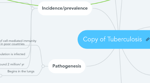
2. Incidence/prevalence
2.1. Over 1.5 million cases/ yr
2.2. Highest incidence
2.2.1. Hospital employees
2.2.2. Drug/alcohol abusers
2.2.3. Homeless
2.2.4. Nursing home residents
2.2.5. Being incarcerated
2.2.6. Elderly men
2.2.7. Children under 15
2.2.8. Hispanic/ Asian populations
2.3. Commonly occur in poor countries
2.4. 40% of the world's population is infected
2.5. Kills around 2 million/ yr
3. Pathogenesis
3.1. Progression depends on number of M. Tuberculosis inhaled, virulence, and development of cell-mediated immunity
3.2. Begins in the lungs
3.2.1. Airborne disease
3.2.1.1. Inhaled bacilli attach to the middle section of the lobes
3.2.1.1.1. 1-1.5 cm of grey white inflammation forms (Ghon focus)
3.2.1.1.2. M. Tuberculosis enters macrophages by endocytosis
3.2.1.1.3. Inhibits Ca++ signals
3.2.1.2. Infection remains latent
4. Manifestations
4.1. Cough
4.1.1. All ages
4.2. Hemoptysis
4.2.1. Elderly
4.3. Night sweats
4.3.1. Specifically in children
4.4. Fever
4.4.1. Prolonged and unexplained
4.4.1.1. All ages
4.5. Chest pain
4.5.1. Adults
4.6. Weight loss
4.6.1. All ages
4.7. Anorexia
4.7.1. Elderly
4.8. Muscle wasting
4.8.1. Elderly
4.9. Malaise
4.9.1. All ages
4.10. Fatigue
4.10.1. All ages
4.11. Poor skin turgor
4.11.1. Adults and elderly
4.12. Dry flaky skin
4.12.1. Elderly
4.13. Stridor
4.13.1. Adults
4.14. Tachypnea
4.14.1. All ages
4.15. Crackles in lungs
4.15.1. All ages
4.16. Mental status change
4.16.1. Specifically elderly
4.17. Dysuria
4.17.1. Specific to Genitourinary TB
4.18. Small caseous lesions
4.18.1. Specific to Miliary TB
4.18.1.1. Elderly have no reactive form
5. Sources
5.1. Godfrey, C., Andersen, J., Mngqibisa, R., Scott, L.E., & Conradie, F. (2016). Tuberculosis control. The Lancet, 387(10024), 1157-1158. doi:http://do.doi.org/10.1016/S0140-6736(16)00706-6
5.2. Kranzer, K., Khan, P., Godfrey-Fausset, P., Ayles, H., & Lönnroth, K. (2016). Tuberculosis control. The Lancet, 387(10024), 1159-1160. Doi:http://dx.doi.org/10.1016/S0140-6736(16)00710-8
5.3. Dheda, K., Barry, C.E., Maartens, G. (2016). Tuberculosis. The Lancet, 387(10024), 1211-1226. Doi:http://dx.doi.org/10.1016/S0140-6736(15)00151-8
5.4. Schmidt, C. W. (2008). Linking TB and the Environment: An Overlooked Mitigation Strategy. Environmental Health Perspectives, 116(11), A478–A485.
6. Diagnostics
6.1. Fluorochrome
6.1.1. Acid-fast bacilli sputum
6.1.1.1. Cultured for M tuberculosis
6.1.1.1.1. Positive sputum is green, purple to, yellowish, mucoid, bloody
6.2. Chest x-ray
6.2.1. Show advanced pulmonary tuberculosis
6.2.1.1. Posterior- anterior chest x-ray is standardized view
6.2.1.1.1. Other organs besides lungs can have an x-ray
6.3. Tissue analysis
6.3.1. Small sample of tissue taken from area believed to be infected
6.4. Mantoux test
6.4.1. Common in children
6.4.1.1. Used around the world
6.4.1.1.1. Intradermal injection of 0.1 mm of PPD tuberculin
6.5. Blood cultures
6.5.1. Common in children over 5 y/o
6.5.1.1. Only indicates if person has been infected with Tb bacteria
6.6. HIV testing
6.6.1. HIV is most known risk factor
6.6.1.1. HIV to TB disease is rapid progression
6.6.1.1.1. Adults specifically
6.6.2. ELISA
6.6.2.1. Third and fourth generation HIV tests
6.6.2.1.1. Looks for specific HIV antibodies in bodily fluids
6.7. ELISpot
6.7.1. T-Spot
6.7.1.1. Counts number of antimycobacterial effector T cells and WBCs that produce interferon-gamma in blood
6.7.1.1.1. Rarely used in children
7. Treatments
7.1. Anti-infective (antibiotics) therapy
7.1.1. 6 to 9 months
7.1.2. INH, RIF, PZA, ETB, Streptomycin, BCG
7.1.2.1. BCG is commonly used in children
7.1.2.2. RIF and PZA are commonly used in elderly
7.1.3. Second line medications: cycloserine, ethionamide, capreomycin sulfate
7.2. Intravenous fluids
7.2.1. Given to those with nutritional compromise
7.3. Coughing / deep breathing exercises
7.3.1. Especially for patients with fibrosis and parenchyma destruction
7.4. Promote bed rest
7.4.1. Increases antibodies being able to cure TB
7.5. Mechanical ventilation
7.5.1. Extreme case
7.5.1.1. Used in those with respiratory failure
7.6. Humidity and oxygen
7.6.1. Stabilizes hypoxia
7.6.2. Decrease thickness of secretions
7.7. Emergency intubation
7.7.1. Extreme case
7.7.1.1. Used especially with pulmonary TB
7.8. Total parenteral nutrition
7.8.1. Given to those with nutritional compromise
