

1. PRESSURE CAUSES BLOOD TO MOVE FROM ONE PLACE TO ANOTHER!
1.1. Opening of valves:
1.1.1. AV valves:
1.1.1.1. Opening
1.1.1.1.1. When AV valves open, it is because atrial pressure is exceeding ventricular pressure. Thus the blood flows into the ventrial
1.1.1.2. Closing
1.1.1.2.1. When AV Valves close it is because ventricular pressure is exceeding atrial pressure
1.1.2. Semi lunar valves
1.1.2.1. Opening:
1.1.2.1.1. When the ventricles contract the pressure exceeds atrial pressure, causing the semi-lunar valve to open (the AV valves are shut)
1.1.2.2. Closing:
1.1.2.2.1. As the ventricles relax, aortic and pulmonary pressure exceed ventricular pressure and the semi-lunar valves snap shut.
2. Cardiac Cells
2.1. dependent upon Aerobic Metabolism
2.1.1. From the krebs cycle
2.2. Hypertrophy in response to increased demand of excersice, and high blood preasure
2.2.1. Good growth
2.2.1.1. during exsercise
2.2.2. Bad growth
2.2.2.1. high blood preasure
3. Excitation - Contraction Coupling
3.1. Much like skeletal muscle (t-tubules, troponin, and tropomyosin)
3.2. External Ca++ joins with Ca++ from the sarcoplasmic reticulum to cause overall increase in intracellular Ca++ (to make a larger contraction?)
3.3. ECG:
3.3.1. P Wave: Spread electrical activity, SA node is depolarizing and causing the atria to contract, (ventricles are relaxing
3.3.2. PQ Segment: Corresponds to the plateu phase of atrial contraction potential. Impulse travels to the AV node. Isovolumetric contraction is occuring here
3.3.2.1. Isovolumetric contraction refers to early ventricular systole and occurs when the AV valve is closed and the pressure is building up the ventricals
3.3.2.1.1. Pressure building up in the ventricles is the reason why it occurs
3.3.3. QRS Complex: Represents the spread of depolarization through the A-V bundle, (of His) bundle branches, Purkinje fibers and ventricular musculature
3.3.4. S-T segment: Ventricles are completely depolarized, this is the plataeu phase of ventricular action potentials its position and shape are important in diognosis:
3.3.5. T Wave: Represents repolarization of the ventricular muscle
3.3.5.1. Isovolumetric relaxation:
3.3.6. T-P Segment: The time from the end of the T wave to the beginning of the P wave of the next cycle. This represents the diastolic pause when the entire heart is depolarized. During most of this segment the heart
3.4. There is always a contraction proceeding that area's depolarization
4. The Cardiac Cycle
4.1. One complete cycle cycle consists of atrial systole, atrial diastole, ventricular stosyole and ventricular diastole
4.1.1. Diastole- relaxation
4.1.2. Systole-contraction
4.2. Identical events are happening simultaneously on the right side of the heart. The only difference is that the pressures generated on the right side are much lower than those generated on the left side
4.3. Important terms
4.3.1. End Diastolic Volume (EDV)
4.3.1.1. Volume at the end of filling (?)
4.3.1.2. the amount of blood in the ventricles at the end of atrial systole just prior to ventricular contraction
4.3.1.2.1. usually refers to the ventricles
4.3.2. End Systolic Volume (ESV)
4.3.2.1. any point when the heart is contracting
4.3.2.1.1. usaually we just mean the ventricles
4.3.3. Stroke Volume
4.3.3.1. How much blood is being pumped per beat
4.3.4. Diastole
4.3.4.1. relaxation
4.3.5. Systole
4.3.5.1. contraction
4.4. The Order of Events: Deplarization
4.5. 1) Blood enters the atria (atrial filling) and may continue on into the ventricles
4.6. 2) Atria contracts to force last bit of blood into ventricles (ventricular filling or diastole)
4.6.1. AV- Valve closes, as atrial pressure is now lower than ventricular pressure
4.7. 3) Ventricles undergo isovolumetric contraction
4.7.1. Isovolumetric Contraction is the initialinitial phase of ventricular contraction in which tension and pressure in the ventricle increase, but no blood is pumped or ejected from the heart
4.8. 4) When ventricular pressure exceeds aortic/pumonary atery pressure, semilunar valves open and ejection occurs.
4.8.1. Semi-lunar valve; both the tricuspid and the mitral valves; the valves that allow blood to
4.9. 5) Semilunar vavles close (dub) when ventricular pressure falls below arterial pressure and ventricles undergo isovolumetric relaxation
4.10. 6. When ventricular pressure falls below atrial pressure and blood that has been accumulating in atria pours into ventricles
4.11. Ventricular Filling
4.11.1. 70% of the blood returning to the heart flows directly through the atrium into the ventricle. Atrial contraction ejects the rest.
4.11.1.1. It is significant that most of the ventricular filling occurs during early diastole.During times of rapid heart rate, the duration of diastole is greatly reduced (more so than systole) which decreases the time for ventricular filling. Since most ventricular filling is occurring early in the diastole period, it is not seriously impaired.
4.11.1.1.1. At heart rates greater than 200 bpm diastole is too short to allow adequate ventricle filling and SV falls.
4.12. Representented
5. Pulmonary Gas Exchange and Transport: Its purpose is to purify O2 by exchanging the metabolic waste of the Krebs Cycle: CO2; and receive O2 to support the Kreb's cycle
5.1. Gas Transport: IMPORTANT
5.1.1. alveoli and capillaries exchange and blood at alveolar capillary interface because those interfaces are so thin
5.1.1.1. Hemoglobin = boat
5.1.1.2. Plasma = river
5.1.1.3. River bed = Capillaries
5.1.1.4. 1 Hemoglobin has four seats for
5.1.2. Carbon Monoxide:
5.1.2.1. attaches onto the hemoglobin
5.2. Atmospheric Pressure: at sea level is 760
5.2.1. 79% Nitrogen
5.2.2. 21% Oxygen
5.2.2.1. PO2= 160 mm Hg @sea level
5.2.2.1.1. 21%/760
5.2.2.2. PO2 = 100 in alveoli
5.2.2.3. PO2 = 120 in exhaled airi
5.2.2.4. Question: Why is there more PO2 in expired air than in the alveoli?
5.2.2.4.1. Initially, the air we intake does all reach the alveoli. This O2 becomes a part of what you expire when you exhale
5.2.3. >1% CO2
5.2.3.1. PCO2 = > 1%
5.3. Chemoreceptors
5.3.1. Ventilation rate and depth may be changing depending on the need to regulate thel blood levels of O2 and CO2 and pH (which is tightly controlled)
5.3.1.1. pH levels are allowed to vary between
5.3.2. Central chemoreceptors
5.3.2.1. very sensitive to changes in PCO2
5.3.2.1.1. but not to other inputs
5.3.3. Periphreal chemoreceptors
5.3.3.1. capable of responding to arterial blood loevels of PO2, PCO2, and pH but are not as senitive
5.3.3.1.1. Primarily respond to decreasing arterial PO2 but it must fall below 60 mm HG to iniate a good response
5.3.4. Only sensing plasma not what's on hemoglobin
5.3.4.1. 98% of plasma is on hemoglobin
5.3.4.1.1. which means, that chemoreceptors are unable to detect Carbon Mondoxide since those molecules take the four seats on hemoglobin
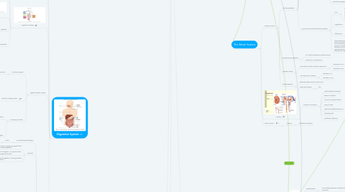
6. Digestive System
6.1. Contribution of other body systems to the digestive system
6.2. The function of the digestive system is to break down the foods you eat, release their nutrients, and absorb those nutrients into the body.
6.3. Digestive Processes
6.3.1. Mastication:
6.3.1.1. Mastication or chewing is the process by which food is crushed and ground by teeth.
6.3.1.1.1. It is the first step of digestion, and it increases the surface area of foods to allow a more efficient break down by enzymes.
6.3.2. Segmentation
6.3.2.1. Segmentation involves contractions of the circular muscles in the digestive tract,
6.3.3. Peristasis
6.3.3.1. hte involuntary constriction and relaxation of the muscles of the intestine or another canal, creating wavelike movements that push the contents of the canal forward.
6.3.4. Digestion
6.3.4.1. actual breakdown of foodstuffs. Digestion can be further categorized as mechanical or chemical digestion
6.3.4.1.1. There are three primary categories of nutrients
6.3.5. absorbtion
6.3.5.1. movement of small "absorbable" molecules from the lumen of the digestive tract to the blood or lymph
6.4. Digestive System Organs
6.4.1. Alimentary Organs
6.4.1.1. "to nourish"
6.4.1.1.1. comprised of
6.4.2. Functions of organ system
6.4.3. Accessory Structures
6.4.3.1. Mechanical digestion
6.4.3.1.1. mouth
6.4.3.1.2. teeth
6.4.3.1.3. tounge
6.4.3.2. Salivary gland
6.4.3.2.1. begins chemical digestion
6.5. Anatomy
6.5.1. Mouth/Pharynx/Esophagus
6.5.1.1. Saliva:
6.5.1.1.1. (a) chemical digestion of carbohydrates
6.5.2. Stomach
6.5.2.1. Major function: storage of ingested food (while awaiting digestion)
6.5.2.2. Chemical digestion: HCI (parirtal cells) Pepsinogen (chief cells)
6.5.2.3. Mechanical digestion = strong peristlatic contractions
6.5.3. Small Intestine; Major function: absorption of nutrients
6.5.3.1. Segments
6.5.3.1.1. duodenum
6.5.3.1.2. jejunum
6.5.3.1.3. Ileum
6.5.3.2. villi and microbilli greatly increase the surface are available for absorbtion
6.5.4. Large instestine or colon: Major function: absorbtion of water and lemination of solid feces
6.5.4.1. segments
6.5.4.1.1. caecum
7. How each of the Autonomic Nervous Systems effect MAP
7.1. Sympathetic
7.1.1. Increases HR:
7.1.2. Increases force of contraction
7.1.3. Increases EDV:
7.1.3.1. by squeezing viens to get more blood into the heart before the ventricular contraction
7.2. Parasympathetic
7.2.1. decreases HR:
7.2.1.1. Bracycardia
8. when a build up of metabolites causes vasodilation to occur so that blood will flow more abudnantly to an area
9. Order of events in the Caridac Cycle: Blood flow and Heart Anatomy
9.1. 1: Deoxygenated blood enters the RA through the vena cava
9.2. 2: Blood goes down to the RV through the tricuspid vavle
9.3. 3: Blood gets pushed through the semi-lunar valve into the pulmonary artery
9.4. 4: Blood goes to the lungs
9.5. 5: From the lungs, oxygenated blood comes back to the heart through the pulmonary vien
9.6. From the pulmonary vein, blood flows into the LA, down the mitral valve and into the LV.
9.7. The strong LV pumps blood into the aorta (through the aortic valve) and to the rest of the body
10. Polarization
10.1. Depolarization
10.1.1. the excitatory response caused by action potentials
10.2. Repolarization
10.2.1. the inhibitory response which causes action potentials to relax
11. Has two types of cells
11.1. Contractile cells
11.1.1. muscle cells -- they do the mechanical work
11.2. Electrical Activity goes praidly to AV node via internodal parth way
11.3. Autorythmic (pacemaker) cells
11.3.1. The Cardiac Conduction Cycle
11.3.1.1. Sympathetic Stimulation
11.3.1.1.1. SA Node
11.3.1.1.2. AV Node
11.3.1.2. SA node Depolarizes
11.3.1.3. Depolarization moves through ventricular conducting system to the apex of the heart
11.3.1.4. Depolarization wave spreads upward from the apex
11.3.2. they are capable of self-generating electrical activity
11.3.2.1. Just like skeletal muscle: Excitation-Contraction coupling: there must be an electrical signal to increase intracellular calcium and start contraction
11.3.2.1.1. The Nervous Systems Role in generating electricity for the heart
11.3.2.2. Parasympathetic Stimulation
11.3.2.2.1. SA Node
11.3.2.2.2. AV Node
12. Blood Preassure and Blood Vessels
12.1. Who gets blood?
12.1.1. Determined By
12.1.1.1. 1) Metabolic need
12.1.1.2. 2) Organ function (ie what is the priority at the moment)
12.1.1.2.1. The brain ALWAYS receives a constant supply of blood.
12.1.2. Caused By: metabolites acting as vasodolaters
12.1.2.1. Technical terms:
12.1.2.1.1. Active Hypernea:
12.1.2.1.2. Reactive Hypernea:
12.2. Blood Vessels
12.2.1. Arteries:
12.2.1.1. take blood away from the heart
12.2.1.1.1. conducting vessels that are PRESSURE (lots of elastic thick walls)
12.2.1.2. Help blood get pumped around the heart
12.2.1.2.1. they have an elastic nature to help push blood around
12.2.1.3. Aretolies
12.2.1.3.1. very small artery that leads to a resistance capillary, ALSO CALLED A RESISTANCE VESSEL
12.2.1.3.2. Sympathetic Nervous System
12.2.2. Capillaries
12.2.2.1. Exchange vessels: walls consist of a single layer of endothelium, smallest of blood vessels where physical exchange occurs between the blood and tissue cells surrounded by interstitial fluid
12.2.2.1.1. Will leak excessive fluid if blood pressure is too high
12.2.3. Veins
12.2.3.1. takes blood too the heart
12.2.3.1.1. capacitance vessles (has a ability to storre electrical charge {probably when blood pressure gets to low])
12.2.3.2. must have vavles because blood pressure is low, so there can't be any backflow
12.3. Liver makes plasma protiens
12.4. MAP = CO X TPR
12.4.1. MAP = Mean Arterial Preassure ( that is average pressure measured in the areterials)
12.4.1.1. CO=Cardiac output HRxSV
12.4.1.1.1. TPR = Resistence
12.5. Blood Pressure Regulation
12.5.1. SHOCK inadequate delivery of blood to tissues
12.5.1.1. HYPOVOLEMIC SHOCK (aka cold shock) inadequate amount of blood (Vasoconstriction)
12.5.1.1.1. Hemorrhagic shock
12.5.1.1.2. Traumatic shock
12.5.1.1.3. Burn Shock
12.5.1.2. CARDIOGENIC OR CIRCULATORY SHOCK
12.5.1.2.1. Myocardial Infarction:
12.5.1.2.2. Congestive Heart Failure
12.5.1.2.3. Arrhythmias:
12.5.2. When MAP drops, the body will quickly attempt to correct the problem
12.5.2.1. MAP is sensed by baroreceptors
12.5.2.1.1. How they work to attempt the problem - Effectors:
12.6. DISTRIBUTIVE SHOCK( aka warm shock) blood volume is normal, but the blood is in the wrong place and is not being delivered to vital tissues (Vasodilation)
12.6.1. Fainting:
12.6.1.1. nerogenic vasodilation of arterioles
12.6.2. Anaphylaxis:
12.6.2.1. systemic (body wide) allergic reaction causing widespread histamine like repsonse
12.6.3. Sepsis
12.6.3.1. reaction to toxins in blood; causes widespread vasodilation of arterioles
12.6.3.1.1. "Tachycardia"
13. Capillaries
14. The Renal System
14.1. Kidney Functions:
14.1.1. Fluid homeostatsis:
14.1.1.1. removes waste products from the body while matinaing a balance of substances for vital body function
14.1.1.1.1. Electrolyte regulation: NA+, K+, H+, Ca2+, Mg3+, C1-, HSO4-, PO3
14.1.1.2. pH levels (have chemoreceptors)
14.1.1.3. MAP
14.1.1.3.1. regulation of salt and chloride ions
14.1.1.4. Osmolarity
14.1.1.4.1. (Angiotensiogen II)
14.1.1.5. Waste Products
14.1.1.5.1. (urea)
14.1.1.6. Receptors involved in fluid homeostasis
14.1.1.6.1. osmoreceptors
14.1.1.6.2. primary baroreceptors
14.1.1.6.3. Renal baroreceptors
14.1.1.7. Hormones involved with fluid homeostasis
14.1.1.7.1. ADH:
14.1.1.7.2. Angiotensin II
14.1.1.7.3. Aldosterone:
14.1.1.7.4. Atrial Natiuretic Peptide (Factor): released from the atria when they are stretched due to increased pressure (increased blood volume). Increases sodium and water excretion, thus reducing blood colume in isosomtic fashion (the opposite of aldosterone) Peptide
14.1.2. Blood pressure regulation
14.1.2.1. NA+ balance (determines blood volume)
14.1.2.2. Production of Angiotensin II
14.1.3. Secretion of EPO
14.1.3.1. stimulates formation of RBS in response to
14.1.3.1.1. decrease of MAP
14.1.3.1.2. decrease in PO2
14.1.3.2. Two ways epo is released
14.1.3.2.1. decrease MAP
14.1.3.2.2. decrease in PO2
14.1.4. Glyconeogenisis
14.1.4.1. synthesis of glucose from amino acids
14.1.4.2. made from kidenys
14.1.4.2.1. liver
14.2. Anatomy
14.2.1. Features of a nephron
14.2.1.1. Afferent/efferent arterolies
14.2.1.2. Bowman's capsule
14.2.1.3. Proximal convoluted tubule
14.2.1.4. Loop of Henle
14.2.1.5. Distal convoluted tubule (DCT)
14.2.1.6. Collecting duct
14.2.1.7. Peritubular capillaries
14.3. Renal Function:
14.3.1. Nephron
14.3.1.1. Peritublular capillaries