1. McCance, K.L., & Huether, S.E. (2014). Pathophysiology: The biologic basis for disease in adults and children, seventh edition. St. Louis, MO: Mosby Elsevier.
2. References:
3. Nazer, D. & Nazer, H. (2017). Pediatric hypertrophic pyloric stenosis. Retrieved from www.emedicine.medscape.com/article/929829-overview
4. Physiologic Etiology
4.1. A disorder that presents shortly after birth
4.2. Caused by narrowing or obstruction of the pylorus secondary to hypertrophy and hyperplasia of the muscular layers of the pylorus
4.3. Approximately 95% of infantile pyloric stenosis are diagnosed in those aged 3-12 weeks
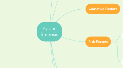
5. Risk Factors
5.1. Suspected environmental factors
5.1.1. Infantile hypergastrinemia
5.1.2. abnormalities in the myenteric plexus innervation
5.1.3. cow's milk protein allergy
5.1.4. exposure to macrolide antibiotics
5.2. Hereditary factors
5.2.1. this condition occurs in as many as 7% of infants of affected parents
6. Common Symptoms
6.1. chronic vomiting
6.2. constipation
6.3. weight loss
6.3.1. Prolonged delay in diagnosis can lead to dehydration, poor weight gain, malnutrition, metabolic alterations, and lethargy.
6.4. electrolyte imbalance
6.5. slight hematemesis
7. Treatments
7.1. this condition can be corrected by surgery
7.1.1. Ramstedt Pyoloromyotomy is the standard procedure of choice
7.1.2. Laparoscopic Pyoloromyotomy may be used
7.1.3. Endoscopic Pyoloromyotomy may also be used and is a simple procedure that can be done outpatient
7.1.4. Endoscopic Balloon Dilatation of hypertorphic pyloric stenosis can be used after failed pyoloromyotomy
8. Genetic Details
8.1. its prevalence is about 3 per 1000 live births in whites
8.2. Much more common in males than females; affects 1 of 200 males and 1 of 1000 females
8.2.1. The difference in prevalence reflects two thresholds in the liability distribution- lower in males and higher in females
8.2.1.1. A lower threshold means that fewer disease-causing factors are required to generate the disorder in males, and females must be exposed to more disease-causing factors to develop the disease
8.2.1.2. Because of this lower threshold, males always have a higher risk than females
8.2.1.3. A family with an affected female must have more genetic and environmental risk factors to produce a higher recurrence risk of the disease in future offspring
8.2.2. The highest risk category would be male relatives of female proband (the individual from which the pedigree begins)
9. Causative Factors
9.1. Multifactoial, hereditary and environmental contributing factors
10. Diagnostic Tests
10.1. Physical Examination
10.1.1. A palpable "olive" lump can be palpated in the right upper quadrant or epigastrium of the abdomen in 60-80% of infants
10.2. Serum electrolytes level
10.3. Ultrasonography
10.4. Barrium Upper GI study
10.4.1. used when ultrasonography is not diagnostic
10.5. Endoscopy
10.5.1. Reserved for patients with atypical clinical signs when ultrasonography and UGI studies are non-diagnostic
