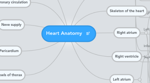
1. Left ventricle
1.1. Openings
1.1.1. Mitral valve
1.1.1.1. Anterior cusp
1.1.1.2. Posterior cusp
1.1.2. Aortic valve
1.1.2.1. Vestibule leads to it (like the infundibulum)
1.1.2.2. right cusp
1.1.2.3. left cusp
1.1.2.4. posterior cusp
1.2. Internal structures
1.2.1. No moderator bands
1.2.2. Two papillary muscles
1.2.3. Thick walls
1.2.4. The lumen is circular in shape
2. Coronary circulation
2.1. Artries
2.1.1. Right coronary artery
2.1.1.1. Sinuatrial nodal branch (60%)
2.1.1.2. Right conus artery
2.1.1.2.1. feeds infundibulum
2.1.1.3. Right marginal branch
2.1.1.3.1. supplies right ventricle
2.1.1.4. Atrioventricular nodal branch (posterior, 65%)
2.1.1.5. Posterior inter ventricular artery (90%, right dominance)
2.1.1.5.1. supplies both ventricles posteriorly
2.1.2. Left coronary artery
2.1.2.1. Anterior inter ventricular artery
2.1.2.1.1. diagonal branch
2.1.2.1.2. left conus artery
2.1.2.1.3. supplies the bundle of HIS
2.1.2.2. Circumflex branch
2.1.2.2.1. Left marginal artery
2.2. Veins
2.2.1. Coronary sinus
2.2.1.1. Small cardiac vein
2.2.1.1.1. with right marginal
2.2.1.2. Middle cardiac vein
2.2.1.2.1. with posterior inter ventricular
2.2.1.3. Great cardiac vein
2.2.1.3.1. with anterior inter ventricular
2.2.2. Right atrium
2.2.2.1. Anterior cardiacl vein
3. Neve supply
3.1. Cardiac plexus
3.1.1. sympathetic
3.1.1.1. preganglionic fibers
3.1.1.1.1. T1-T6
3.1.1.2. Postganglionic fibers
3.1.1.2.1. cervical trunk
3.1.1.2.2. upper thoracic trunk
3.1.1.3. efferent
3.1.1.3.1. increasing rate & contraction force
3.1.1.3.2. vasoconstriction
3.1.1.4. afferent
3.1.1.4.1. referred pain
3.1.2. parasympathetic
3.1.2.1. vagus nerve
3.1.2.2. efferent
3.1.2.2.1. lowering rate & contraction force
3.1.2.2.2. vasoconstriction
3.1.2.3. afferent
3.1.2.3.1. reflexes
4. Pericardium
4.1. surface anatomy
4.1.1. posterior to the sternum, 2-6 costal cartilages
4.1.2. anterior to 5-8 thoracic vertebrae
4.2. Fibrous pericardium
4.2.1. anchors the heart and prevents over stretching
4.2.2. attached to diaphragm, sternum, and major blood vessels
4.2.3. Innervated by the phrenic nerve
4.3. Serous pericardium
4.3.1. parietal layer -> fused to the fibrous pericardium
4.3.1.1. innervated by phrenic nerve
4.3.2. visceral layer (epicardium)
4.3.2.1. innervated by sympathetic trunk & vagus (it's part of the heart)
4.3.3. both are continues around major blood vessles
4.3.4. 50 ml of pericardial fluid in the cavity between them
4.4. Pericardial sinuses
4.4.1. result during development and has NO clinical significance
4.4.2. Transverse sinus
4.4.2.1. between arteries and veins
4.4.3. Oblique sinus
4.4.3.1. within the veins
5. Large vessels of thorax
5.1. Artries
5.1.1. Ascending aorta
5.1.1.1. it's inside the pericardium
5.1.1.2. right and left coronary arteries
5.1.2. Arch of aorta
5.1.2.1. Brachiocephalic trunk
5.1.2.1.1. Right subclavian
5.1.2.1.2. Right common carotid
5.1.2.1.3. behind the right sternoclavicular joint
5.1.2.1.4. right recurrent laryngeal nerve
5.1.2.2. left common carotid
5.1.2.3. left subclavian
5.1.2.4. ligamentum arteriosum binds it with pulmonary trunk biforcation
5.1.2.5. left recurrent laryngeal nerve encircles it
5.1.3. Descending thoracic aorta
5.1.3.1. Posterior intercostals
5.1.3.2. subcostals
5.1.3.3. pericardial
5.1.3.4. oesophageal
5.1.3.5. bronchial
5.1.4. Pulmonary trunk
5.1.4.1. Right pulmonary artery
5.1.4.1.1. goes behind ascending aorta
5.1.4.2. Left pulmonary artery
5.1.4.2.1. goes in front of descending aorta
5.1.4.3. also inside the pericardium with the ascending aorta
5.2. Veins
5.2.1. Superior Vena Cava
5.2.1.1. left brachiocephalic
5.2.1.2. right brachiocephalic
5.2.1.2.1. right subclavian
5.2.1.2.2. internal jaguar
5.2.1.2.3. forms at the root of the neck
5.2.2. Azygus veins
5.2.2.1. Main Azygus vein
5.2.2.1.1. most anterior
5.2.2.1.2. on the right side
5.2.2.1.3. ascends though aortic opening of diaphragm at 5th thoracic vertebra
5.2.2.1.4. superior hemiazygos
5.2.2.1.5. Inferior hemiazygos (Accessory)
5.2.2.1.6. right subcostal
5.2.2.1.7. right ascending lumber
5.2.3. Inferior Vena Cava
5.2.4. Pulmonary veins
6. Right atrium
6.1. Posterior wall
6.1.1. Smooth
6.1.2. It is the inter atrial septum
6.1.2.1. Fossa ovalis
6.1.2.2. Anulus ovalis
6.1.2.3. Atrioventricular (AV) node
6.1.2.3.1. most anterior inferior
6.1.2.3.2. septal cusp of tricuspid
6.1.3. 3 openings
6.1.3.1. Superior vena cava
6.1.3.1.1. NO valve
6.1.3.2. Inferior vena cava
6.1.3.2.1. Rudimentary valve
6.1.3.3. coronary sinus orifice
6.1.3.3.1. between the inferior vena cava and the right AV orifice
6.1.3.3.2. Rudimentary valve
6.2. Anterior wall
6.2.1. Rough -> musculi pectinati
6.2.2. Right auricle
6.2.2.1. what separates if from the posterior wall
6.2.2.1.1. Sulcus terminalis (external)
6.2.2.1.2. Crista terminalis (internal)
6.3. Right AV valve (tricuspid)
7. Right ventricle
7.1. Openings
7.1.1. Tricuspid valve
7.1.1.1. anterior cusp
7.1.1.2. septal cusp
7.1.1.3. posterior (inferior) cusp
7.1.2. Pulmonary valve
7.1.2.1. A funnel shaped structure leads to it (the infundibulum)
7.1.2.2. right cusp
7.1.2.3. left cusp
7.1.2.4. anterior cusp
7.2. Internal structures
7.2.1. Trabeculae carneae
7.2.1.1. ridges of cardiac muscles
7.2.1.2. 3 papillary muscles, each gives chordae tendineae to 2 cusps
7.2.1.3. moderator band
7.2.1.3.1. Shortcut to convoy the right branch of the AV bundle
7.2.2. Inter ventricular wall
7.2.2.1. Membranous wall
7.2.2.1.1. very thin, superior
7.2.2.2. Muscular part
7.2.3. Lumen is crescent shape
8. Orientation
8.1. Apex
8.1.1. left ventricle
8.2. Base
8.2.1. Mainly left atrium
8.2.2. right atrium
8.3. Anterior surface
8.3.1. Mainly right ventricle
8.3.2. right atrium
8.3.3. left ventricle
8.4. Inferior surface
8.4.1. Mainly left ventricle
8.4.2. Right ventricle
8.5. Right border
8.5.1. Right atrium
8.6. Left border
8.6.1. Left ventricle
8.6.2. Left auricle
8.7. Inferior border
8.7.1. Right ventricle
8.8. Superior border
8.8.1. Vessels
9. Skeleton of the heart
9.1. Fibrous rings surrounding the heart, made of collagen
9.2. Fibrous trigons connect the rings
9.3. Make up the membranous inter ventricular septum
9.4. Function
9.4.1. electrical insulation
9.4.2. support and muscle attachment
10. Left atrium
10.1. similar structure to the right atirum
10.2. Openings
10.2.1. 4 pulmonary viens
10.2.1.1. Left superior pulmonary vien
10.2.1.2. Left inferior pulmonary vien
10.2.1.3. 2 right pulmonary viens
10.2.1.4. NO valves
10.2.2. Mitral valve (left AV)

