

1. Reticular Tissue & Adipose Tissue
1.1. Reticular Tissue
1.1.1. Structure
1.1.1.1. Reticular Fibers:
1.1.1.1.1. These fibers form a fine, mesh-like network (reticulum) that supports the structure of various organs.
1.1.1.1.2. Composed primarily of Type III collagen.
1.1.1.1.3. Reticular fibers are heavily glycosylated, giving them an affinity for silver stains, where they appear black (argyrophilic).
1.1.1.2. Cells:
1.1.1.2.1. Reticular Cells:
1.1.1.2.2. Other Cells:
1.1.1.3. Ground Substance:
1.1.1.3.1. The ground substance in reticular tissue is sparse but contains glycoproteins and proteoglycans that help maintain the structure of the reticular fibers.
1.1.2. Function
1.1.2.1. Structural Support:
1.1.2.1.1. Provides a delicate, supportive framework for soft tissues, especially in hematopoietic (blood-forming) and lymphatic organs.
1.1.2.2. Filtration:
1.1.2.2.1. In organs like the spleen and lymph nodes, reticular tissue acts as a filter, trapping foreign particles and facilitating the immune response.
1.1.2.3. Cellular Organization:
1.1.2.3.1. Reticular tissue organizes and maintains the spatial arrangement of cells in tissues like the bone marrow, spleen, and lymph nodes.
1.1.3. Locations
1.1.3.1. Lymphoid Organs:
1.1.3.1.1. Found in lymph nodes, spleen, and tonsils, where it supports the immune cells.
1.1.3.2. Bone Marrow:
1.1.3.2.1. Provides the structural framework for hematopoietic stem cells and developing blood cells.
1.1.3.3. Liver:
1.1.3.3.1. In the liver, reticular fibers form the stroma that supports hepatocytes and sinusoids.
1.1.4. Histological Appearance
1.1.4.1. Light Microscopy:
1.1.4.1.1. Reticular fibers are not easily seen with Hematoxylin and Eosin (H&E) staining due to their thinness.
1.1.5. Pathological Conditions
1.1.5.1. Reticulosis:
1.1.5.1.1. An abnormal increase in reticular fibers, often seen in chronic inflammation or certain tumors.
1.1.5.2. Reticular Dysplasia:
1.1.5.2.1. A rare condition where reticular tissue is underdeveloped, affecting the function of the affected organ, particularly in the immune system.
1.2. Adipose Tissue
1.2.1. Structure
1.2.1.1. Adipocytes (Fat Cells):
1.2.1.1.1. The primary cell type in adipose tissue.
1.2.1.2. Fibers:
1.2.1.2.1. Reticular Fibers:
1.2.1.2.2. Collagen Fibers:
1.2.1.3. Vascularization:
1.2.1.3.1. Adipose tissue is highly vascularized, ensuring an adequate supply of nutrients and oxygen to the adipocytes and facilitating the release of fatty acids into the bloodstream.
1.2.1.4. Ground Substance:
1.2.1.4.1. Sparse, consisting mainly of proteoglycans and glycoproteins that provide minimal structural support.
1.2.2. Types of Adipose Tissue
1.2.2.1. White Adipose Tissue (WAT):
1.2.2.1.1. Locations:
1.2.2.1.2. Function:
1.2.2.2. Brown Adipose Tissue (BAT):
1.2.2.2.1. Function:
1.2.2.2.2. Locations:
1.2.3. Histological Appearance
1.2.3.1. White Adipose Tissue:
1.2.3.1.1. Light Microscopy:
1.2.3.2. Brown Adipose Tissue:
1.2.3.2.1. Light Microscopy:
1.2.4. Functions
1.2.4.1. White Adipose Tissue:
1.2.4.1.1. Thermal Insulation: Prevents heat loss by forming an insulating layer under the skin.
1.2.4.1.2. Energy Storage: Stores triglycerides as an energy reserve that can be mobilized during fasting or increased energy demand.
1.2.4.1.3. Mechanical Cushioning: Protects organs by absorbing shock and pressure.
1.2.4.1.4. Endocrine Regulation: Produces hormones that regulate appetite, metabolism, and insulin sensitivity.
1.2.4.2. Brown Adipose Tissue:
1.2.4.2.1. Heat Production: Generates heat, especially in newborns and hibernating animals, to maintain body temperature in cold environments.
1.2.4.2.2. Energy Expenditure: Plays a role in burning calories and may contribute to weight regulation
1.2.5. Pathological Conditions
1.2.5.1. Obesity:
1.2.5.1.1. Excessive accumulation of white adipose tissue, leading to increased body weight and associated metabolic disorders such as insulin resistance, type 2 diabetes, and cardiovascular disease.
1.2.5.2. Lipomas:
1.2.5.2.1. Benign tumors composed of mature white adipose tissue, usually soft and painless, occurring in various parts of the body.
2. Loose and Dense Connective Tissue
2.1. Loose Connective Tissue
2.1.1. Structure:
2.1.1.1. Fibers:
2.1.1.1.1. Elastic Fibers: These thin, branching fibers are present in small amounts and allow the tissue to stretch and then return to its original shape.
2.1.1.1.2. Collagen Fibers: These are the most abundant fibers in loose connective tissue, providing some tensile strength while allowing flexibility. They are loosely packed and run in various directions, giving the tissue a less organized appearance.
2.1.1.1.3. Reticular Fibers: Form a fine meshwork that supports the structure of various organs, especially in the stroma of soft tissues like the liver and lymph nodes.
2.1.1.2. Cells:
2.1.1.2.1. Mast Cells: Contain granules rich in histamine and heparin, playing a role in inflammatory responses.
2.1.1.2.2. Fibroblasts: The most common cells in loose connective tissue, responsible for synthesizing collagen, elastic fibers, and ground substance.
2.1.1.2.3. Macrophages: Immune cells involved in phagocytosis and defense against pathogens.
2.1.1.2.4. Adipocytes: Fat cells that store energy and provide insulation.
2.1.1.2.5. Plasma Cells: Derived from B lymphocytes, they produce antibodies.
2.1.1.2.6. Other Immune Cells: Including lymphocytes, neutrophils, and eosinophils, which participate in immune responses.
2.1.1.3. Ground Substance:
2.1.1.3.1. Composed of glycosaminoglycans (GAGs), proteoglycans, and glycoproteins.
2.1.1.3.2. The ground substance is a gel-like matrix that fills the space between fibers and cells, allowing for the diffusion of nutrients, gases, and waste products.
2.1.2. Functions:
2.1.2.1. Support and Binding:
2.1.2.1.1. Loose connective tissue binds organs and tissues together, providing structural support without restricting movement.
2.1.2.2. Nutrient Supply:
2.1.2.2.1. The ground substance serves as a medium for the exchange of nutrients and waste products between blood vessels and surrounding cells.
2.1.2.3. Immune Defense:
2.1.2.3.1. Contains immune cells that protect against infection and participate in inflammatory responses.
2.1.2.4. Flexibility:
2.1.2.4.1. The loose arrangement of fibers allows the tissue to stretch, bend, and return to its original shape without damage.
2.1.3. Locations:
2.1.3.1. Underneath Epithelia: Loose connective tissue forms the lamina propria, supporting epithelia in the digestive, respiratory, and urinary tracts.
2.1.3.2. Around Blood Vessels and Nerves: Provides support and protection.
2.1.3.3. In Serous Membranes: Found in pleura, pericardium, and peritoneum, supporting mesothelial cells and allowing smooth gliding of organs.
2.1.3.4. Filling Spaces Between Organs: Helps maintain the position of organs within body cavities.
2.1.4. Histological Appearance:
2.1.4.1. Light Microscopy:
2.1.4.1.1. The ground substance is not stained and appears as empty spaces between the fibers and cells.
2.1.4.1.2. Under H&E staining, loose connective tissue appears as a loosely organized matrix with thin, wavy collagen fibers, scattered elastic fibers, and a variety of cell types.
2.2. Dense Connective Tissue
2.2.1. Structure:
2.2.1.1. Fibers:
2.2.1.1.1. Elastic Fibers: Present in varying amounts, especially in dense elastic tissue, contributing to the tissue’s ability to stretch and recoil.
2.2.1.1.2. Collagen Fibers: Predominate in dense connective tissue, arranged in either parallel (dense regular) or irregular (dense irregular) patterns, depending on the tissue’s specific function.
2.2.1.2. Cells:
2.2.1.2.1. Fibroblasts: The primary cell type, responsible for the production and maintenance of the dense collagen fibers.
2.2.1.2.2. Fewer Immune Cells: Compared to loose connective tissue, dense connective tissue contains fewer immune cells, as its primary role is structural support rather than immune defense.
2.2.1.3. Ground Substance:
2.2.1.3.1. There is relatively little ground substance compared to loose connective tissue, as the space between fibers is minimal.
2.2.2. Types of Dense Connective Tissue:
2.2.2.1. Dense Regular Connective Tissue:
2.2.2.1.1. Structure:
2.2.2.1.2. Function:
2.2.2.1.3. Locations:
2.2.2.1.4. Histological Appearance:
2.2.2.2. Dense Irregular Connective Tissue:
2.2.2.2.1. Structure:
2.2.2.2.2. Function:
2.2.2.2.3. Locations:
2.2.2.2.4. Histological Appearance:
2.2.2.3. Dense Elastic Connective Tissue:
2.2.2.3.1. Structure:
2.2.2.3.2. Function:
2.2.2.3.3. Locations:
2.2.2.3.4. Histological Appearance:
2.3. Functional Comparison
2.3.1. Loose Connective Tissue:
2.3.1.1. Vascularity: Typically more vascular, facilitating the exchange of nutrients and waste between blood and cells.
2.3.1.2. Flexibility: Provides more flexibility and less tensile strength, suitable for supporting epithelia, cushioning organs, and allowing the passage of immune cells.
2.3.2. Dense Connective Tissue:
2.3.2.1. Strength: Provides greater tensile strength and resistance to stretching, suitable for tendons, ligaments, and organ capsules.
2.3.2.2. Vascularity: Generally less vascular than loose connective tissue, especially in dense regular connective tissue, which makes healing slower in these tissues.
2.4. Pathological Conditions Related to Connective Tissue
2.4.1. Edema:
2.4.1.1. Accumulation of excess fluid in loose connective tissue, often due to inflammation or impaired lymphatic drainage, leading to swelling.
2.4.2. Tendinitis:
2.4.2.1. Inflammation of tendons, often due to overuse or injury, affecting dense regular connective tissue.
2.4.3. Fibrosis:
2.4.3.1. Excessive deposition of collagen fibers in response to chronic injury or inflammation, leading to stiffening and scarring of tissues, such as in pulmonary fibrosis.
3. Connective Tissue Fibers
3.1. Collagen Fibers
3.1.1. Structure:
3.1.1.1. Basic Composition:
3.1.1.1.1. Collagen molecules are triple helices formed by three polypeptide chains (α-chains), which are rich in glycine, proline, and hydroxyproline.
3.1.1.1.2. Collagen fibers are composed of the protein collagen, which is the most abundant protein in the human body.
3.1.1.1.3. These helices assemble into fibrils, which then bundle together to form fibers.
3.1.1.2. Types of Collagen:
3.1.1.2.1. Type III Collagen: Forms reticular fibers, found in the liver, spleen, and lymphatic tissues. It provides a supportive mesh.
3.1.1.2.2. Type I Collagen: The most common type, found in tendons, ligaments, skin, bone, and most connective tissues. It provides tensile strength.
3.1.1.2.3. Type II Collagen: Predominantly found in cartilage. It forms fine fibrils and provides resistance to pressure.
3.1.1.2.4. Type IV Collagen: Found in the basal lamina of the basement membrane, providing structural support.
3.1.1.2.5. Type V and others: Associated with collagen fibers and basement membranes, playing various roles in tissue structure and function.
3.1.2. Function:
3.1.2.1. Tensile Strength:
3.1.2.1.1. Collagen fibers provide significant tensile strength to connective tissues, allowing them to resist stretching and tearing forces.
3.1.2.2. Structural Support:
3.1.2.2.1. They form the bulk of connective tissue in skin, tendons, ligaments, and bone, providing structural integrity to these tissues.
3.1.2.3. Wound Healing:
3.1.2.3.1. Collagen is essential in wound healing, where fibroblasts produce collagen to form a new extracellular matrix that repairs damaged tissue.
3.1.3. Histological appearence:
3.1.3.1. Light Microscopy:
3.1.3.1.1. Collagen fibers appear as thick, wavy, eosinophilic (pink) fibers when stained with Hematoxylin and Eosin (H&E).
3.1.3.1.2. They are arranged in parallel bundles in tendons and ligaments, providing strength in one direction.
3.1.3.1.3. In loose connective tissue, collagen fibers are more randomly arranged.
3.2. Elastic Fibers
3.2.1. Structure:
3.2.1.1. Basic Composition:
3.2.1.1.1. Elastin is surrounded by a network of microfibrils composed of the glycoprotein fibrillin, which provides a scaffold for elastin deposition.
3.2.1.1.2. Elastic fibers are composed of the protein elastin, which is rich in glycine and proline, like collagen, but contains fewer hydroxyproline residues.
3.2.1.1.3. The fibers can stretch up to 1.5 times their length and return to their original shape, giving tissues elasticity.
3.2.2. Function:
3.2.2.1. Elasticity:
3.2.2.1.1. Elastic fibers allow tissues to stretch and then return to their original shape. This property is crucial in tissues that undergo repeated deformation, such as the skin, lungs, and blood vessels.
3.2.2.2. Resilience:
3.2.2.2.1. They provide resilience to tissues, enabling them to withstand mechanical stresses and maintain their structure over time.
3.2.3. Histological appearence:
3.2.3.1. Light Microscopy:
3.2.3.1.1. They are usually difficult to distinguish with H&E staining due to their refractile nature.
3.2.3.1.2. Elastic fibers are thinner than collagen fibers and often appear as fine, branching fibers.
3.3. Reticular Fibers
3.3.1. Structure:
3.3.1.1. Basic Composition:
3.3.1.1.1. They form a fine, branching network rather than thick bundles, creating a delicate framework in various organs.
3.3.1.1.2. Reticular fibers are composed mainly of Type III collagen.
3.3.1.1.3. These fibers are associated with glycoproteins, which make them more heavily glycosylated than other collagen fibers.
3.3.2. Function:
3.3.2.1. Supportive Framework:
3.3.2.1.1. They provide structural support while allowing the passage of cells and fluids within these tissues.
3.3.2.1.2. Reticular fibers form a supportive network in soft tissues such as the liver, lymph nodes, spleen, and bone marrow.
3.3.2.2. Scaffolding:
3.3.2.2.1. Reticular fibers act as scaffolding for parenchymal cells in organs, maintaining the structure and organization of the tissue.
3.3.3. Histological appearence:
3.3.3.1. Light Microscopy:
3.3.3.1.1. Reticular fibers are not well visualized with H&E staining due to their fine structure.
3.4. Functional Relationships and Distribution
3.4.1. Tendons and Ligaments:
3.4.1.1. Collagen fibers predominate, providing high tensile strength for attaching muscles to bones (tendons) or linking bones together (ligaments).
3.4.2. Skin (Dermis):
3.4.2.1. The dermis contains both collagen and elastic fibers, with collagen providing strength and elasticity allowing the skin to stretch and recoil.
3.4.3. Arteries:
3.4.3.1. Elastic fibers are abundant in the walls of large arteries like the aorta, where they allow the vessel to stretch during blood ejection and then return to its original shape to maintain blood pressure.
3.4.4. Lymphatic Organs:
3.4.4.1. Reticular fibers form the supportive stroma in lymphatic organs such as lymph nodes and the spleen, creating a mesh-like structure that supports immune cells.
4. Ground Substance in Connective Tissue
4.1. Composition of Ground Substance
4.1.1. Glycosaminoglycans (GAGs)
4.1.1.1. Structure:
4.1.1.1.1. GAGs are long, unbranched polysaccharide chains composed of repeating disaccharide units.
4.1.1.1.2. These units usually consist of a sugar acid (e.g., glucuronic acid) and an amino sugar (e.g., N-acetylglucosamine or N-acetylgalactosamine).
4.1.1.1.3. GAGs are highly negatively charged due to the presence of sulfate and carboxyl groups, which attract water molecules, giving the ground substance its gel-like consistency.
4.1.1.2. Types:
4.1.1.2.1. Hyaluronic Acid:
4.1.1.2.2. Chondroitin Sulfate:
4.1.1.2.3. Dermatan Sulfate:
4.1.1.2.4. Heparan Sulfate:
4.1.1.2.5. Keratan Sulfate:
4.1.1.2.6. Heparin:
4.1.2. Proteoglycans
4.1.2.1. Structure:
4.1.2.1.1. Proteoglycans consist of a core protein to which one or more GAG chains are covalently attached.
4.1.2.1.2. The core protein is synthesized in the rough endoplasmic reticulum, and the GAG chains are added in the Golgi apparatus.
4.1.2.2. Function:
4.1.2.2.1. Proteoglycans provide structural support to the ECM by forming a hydrated gel that resists compressive forces.
4.1.2.2.2. They interact with other ECM components, such as collagen and elastin fibers, to maintain tissue integrity.
4.1.2.2.3. Proteoglycans also play a role in cell signaling, influencing cell growth, differentiation, and migration
4.1.3. Multiadhesive Glycoproteins
4.1.3.1. Structure:
4.1.3.1.1. Multiadhesive glycoproteins are proteins with attached carbohydrate chains.
4.1.3.1.2. These molecules have multiple binding sites that allow them to interact with cells, collagen fibers, and proteoglycans.
4.1.3.2. Function:
4.1.3.2.1. They play a key role in stabilizing the ECM and linking it to the cell surface.
4.1.3.2.2. Involved in cell migration, adhesion, proliferation, and differentiation.
4.2. Functions of Ground Substance
4.2.1. Hydration and Diffusion:
4.2.1.1. The ground substance is highly hydrated due to its GAG content, allowing it to resist compressive forces and act as a medium for the diffusion of nutrients, gases, and waste products between blood vessels and cells.
4.2.2. Lubrication:
4.2.2.1. In joints and other tissues, the ground substance provides lubrication, reducing friction between moving parts.
4.2.3. Barrier Function:
4.2.3.1. The ground substance acts as a physical barrier to the movement of pathogens and large molecules, contributing to tissue protection.
4.2.4. Cellular Signaling:
4.2.4.1. Proteoglycans and glycoproteins in the ground substance interact with cell surface receptors, influencing cell behavior, including growth, migration, and differentiation.
4.2.5. Tissue Repair and Regeneration:
4.2.5.1. During tissue repair, the ground substance is remodeled, facilitating the migration of fibroblasts and other cells to the site of injury.
5. Connective Tissue Cells
5.1. Classification of Connective Tissue Cells
5.1.1. Fixed (Resident) Cells
5.1.1.1. Definition:
5.1.1.1.1. These cells are permanent residents of the connective tissue and are primarily involved in the production and maintenance of the ECM.
5.1.1.2. Types:
5.1.1.2.1. 1. Fibroblasts
5.1.1.2.2. 2. Adipocytes (Fat Cells)
5.1.1.2.3. Pericytes
5.1.1.2.4. Mast Cells
5.1.1.2.5. Macrophages (Histiocytes)
5.1.2. Transient (Wandering) Cells
5.1.2.1. Definition:
5.1.2.1.1. These cells migrate into the connective tissue from the bloodstream, especially during immune responses or inflammation.
5.1.2.2. Types:
5.1.2.2.1. Lymphocytes
5.1.2.2.2. Plasma Cells
5.1.2.2.3. Neutrophils
5.1.2.2.4. Eosinophils
5.1.2.2.5. Basophils
5.1.2.2.6. Monocytes
5.2. Role of Connective Tissue Cells in ECM Production
5.2.1. Fibroblasts
5.2.1.1. are the principal cells responsible for synthesizing the components of the extracellular matrix:
5.2.1.1.1. Collagen Fibers:
5.2.1.1.2. Elastin Fibers:
5.2.1.1.3. Reticular Fibers:
5.2.1.1.4. Ground Substance:
5.3. Specialized Connective Tissue Cells
5.3.1. Adipocytes:
5.3.1.1. Store and metabolize fats, with white adipocytes primarily involved in energy storage and brown adipocytes in thermogenesis.
5.3.2. Chondrocytes:
5.3.2.1. Found in cartilage, they maintain the cartilaginous matrix composed of collagen, elastin, and proteoglycans.
5.3.3. Osteoblasts and Osteocytes:
5.3.3.1. Osteoblasts produce the bone matrix, which is mineralized with calcium salts.
5.3.3.2. Osteocytes are mature bone cells embedded in the matrix they helped create.
6. Functions of Glandular Epithelia
6.1. Secretion:
6.1.1. Endocrine glands secrete hormones that regulate metabolism, growth, and other essential bodily functions.
6.1.2. Exocrine glands produce enzymes, mucus, sweat, oil, and other substances vital for digestion, lubrication, and protection.
6.2. Absorption and Modification:
6.2.1. Some exocrine glands (e.g., salivary glands) not only secrete but also modify the ionic composition of their secretions.
6.2.2. Duct cells play a key role in absorbing or adding electrolytes to the secretions.
6.3. Protection:
6.3.1. Glandular epithelia protect underlying tissues by producing mucus (which traps pathogens and debris) and secreting enzymes that digest food particles or inactivate harmful agents.
7. Glandular Epithelia
7.1. Classification of Glands
7.1.1. Exocrine Glands
7.1.1.1. Definition
7.1.1.1.1. Exocrine glands secrete their products onto a surface or into a body cavity through ducts.
7.1.1.2. Classification Based on Duct Structure:
7.1.1.2.1. Simple Glands
7.1.1.2.2. Compound Glands
7.1.1.3. Classification Based on Mode of Secretion:
7.1.1.3.1. Merocrine Secretion
7.1.1.3.2. Apocrine Secretion
7.1.1.3.3. Holocrine Secretion
7.1.1.4. Classification Based on the Nature of Secretion:
7.1.1.4.1. Serous Glands
7.1.1.4.2. Mucous Glands
7.1.1.4.3. Mixed (Seromucous) Glands
7.1.2. Endocrine Glands
7.1.2.1. Definition
7.1.2.1.1. Endocrine glands are ductless glands that release hormones directly into the bloodstream.
7.1.2.2. Structure
7.1.2.2.1. Cord and Clump Type: Cells are arranged in cords or clusters around capillaries or sinusoids.
7.1.2.2.2. Follicular Type: Cells are arranged in follicles, with secretions stored within the follicle lumen.
7.1.2.3. Functions
7.1.2.3.1. Secrete hormones that regulate various physiological processes such as growth, metabolism, and reproduction.
7.2. Histological Features of Glandular Epithelia
7.2.1. Exocrine Glands
7.2.1.1. Acinus (Secretory Unit):
7.2.1.1.1. The basic secretory unit of exocrine glands is the acinus (or alveolus), a cluster of cells that produce the gland's secretions.
7.2.1.2. Duct System:
7.2.1.2.1. Intercalated Ducts
7.2.1.2.2. Striated Ducts
7.2.1.2.3. Excretory Ducts
7.2.1.3. Myoepithelial Cells:
7.2.1.3.1. Specialized contractile cells found around the acini and ducts of some exocrine glands.
7.2.1.3.2. They help expel secretions from the acini into the ducts.
7.2.2. Endocrine Glands
7.2.2.1. Cell Arrangement:
7.2.2.1.1. Cords and Clusters:
7.2.2.1.2. Follicles:
7.2.2.2. Secretion and Storage:
7.2.2.2.1. Endocrine cells produce hormones that are either stored intracellularly (in vesicles) or extracellularly (in follicles, like in the thyroid).
7.2.2.2.2. Hormones are released into the surrounding capillaries and then enter the bloodstream.
7.3. Functions of Glandular Epithelia
7.3.1. Secretion:
7.3.1.1. Endocrine glands secrete hormones that regulate metabolism, growth, and other essential bodily functions.
7.3.1.2. Exocrine glands produce enzymes, mucus, sweat, oil, and other substances vital for digestion, lubrication, and protection.
7.3.2. Absorption and Modification:
7.3.2.1. Some exocrine glands (e.g., salivary glands) not only secrete but also modify the ionic composition of their secretions.
7.3.2.2. Duct cells play a key role in absorbing or adding electrolytes to the secretions.
7.3.3. Protection:
7.3.3.1. Glandular epithelia protect underlying tissues by producing mucus (which traps pathogens and debris) and secreting enzymes that digest food particles or inactivate harmful agents.
8. Functions of Stratified Epithelia
8.1. Protection:
8.1.1. Stratified epithelia provide robust protection against mechanical stress, pathogens, and dehydration.
8.1.2. Keratinized stratified squamous epithelium is particularly effective at preventing water loss.
8.2. Secretion:
8.2.1. Pseudostratified epithelia are often involved in mucus secretion, particularly in the respiratory tract.
8.2.2. Goblet cells within pseudostratified epithelia produce mucus to trap debris and pathogens.
8.3. Stretching and Distension:
8.3.1. Transitional epithelium allows organs like the bladder to stretch as they fill with urine.
8.4. Ciliary Movement:
8.4.1. In the respiratory tract, pseudostratified ciliated columnar epithelium helps clear mucus and foreign particles.
9. Stratified Epithelia
9.1. Stratified Squamous Epithelium
9.1.1. Structure:
9.1.1.1. Multiple layers of cells, with the basal cells typically cuboidal or columnar, and the surface cells being squamous (flat).
9.1.1.2. The basal layer is mitotically active, producing new cells that migrate towards the surface.
9.1.2. Subtypes:
9.1.2.1. Keratinized Stratified Squamous Epithelium:
9.1.2.1.1. Surface cells are dead and filled with keratin, a tough, fibrous protein.
9.1.2.1.2. Provides a waterproof barrier and protection against abrasion.
9.1.2.1.3. Locations: Skin (epidermis).
9.1.2.2. Non-Keratinized Stratified Squamous Epithelium:
9.1.2.2.1. Surface cells are alive, moist, and do not contain keratin.
9.1.2.2.2. Provides protection while remaining pliable.
9.1.2.2.3. Locations: Oral cavity, esophagus, vagina, and cornea.
9.1.3. Functions:
9.1.3.1. Protection against mechanical and chemical damage.
9.1.3.2. Barrier against pathogens.
9.1.3.3. Prevents dehydration (in keratinized types).
9.2. Stratified Cuboidal Epithelium
9.2.1. Structure:
9.2.1.1. Usually two to three layers of cuboidal cells.
9.2.1.2. Less common compared to other stratified epithelia.
9.2.2. Functions:
9.2.2.1. Provides protection and structural support.
9.2.2.2. Involved in secretion and absorption.
9.2.3. Loactions:
9.2.3.1. Ducts of sweat glands, salivary glands, and mammary glands.
9.2.3.2. Developing ovarian follicles.
9.3. Stratified Columnar Epithelium
9.3.1. Structure:
9.3.1.1. Multiple layers, with the surface cells being columnar and the deeper layers more cuboidal.
9.3.1.2. Rarely found in the body.
9.3.2. Functions:
9.3.2.1. Protection and secretion.
9.3.3. Loactions:
9.3.3.1. Parts of the pharynx, male urethra, and large ducts of some glands (e.g., parotid gland).
9.4. Transitional Epithelium (Urothelium)
9.4.1. Structure:
9.4.1.1. Multiple layers of cells that can stretch and contract.
9.4.1.2. Surface cells are dome-shaped or umbrella-shaped when relaxed and flatten when stretched.
9.4.1.3. The basal layer is cuboidal or columnar, and the intermediate layers are more polygonal.
9.4.2. Functions:
9.4.2.1. Allows for stretching and distension without damage.
9.4.2.2. Protects underlying tissues from the toxic effects of urine.
9.4.3. Loactions:
9.4.3.1. Lining of the urinary bladder, ureters, and part of the urethra.
9.5. Pseudostratified Epithelium
9.5.1. Structure:
9.5.1.1. Single layer of cells with varying heights.
9.5.1.2. Nuclei are at different levels, giving the appearance of stratification.
9.5.1.3. Often contains cilia and goblet cells (mucus-secreting).
9.5.2. Functions:
9.5.2.1. Secretion (mainly mucus).
9.5.2.2. Ciliary movement helps in the propulsion of mucus and trapped particles.
9.5.3. Loactions:
9.5.3.1. Respiratory Tract:
9.5.3.1.1. Trachea, bronchi, and nasal cavity (referred to as respiratory epithelium).
9.5.3.2. Male Reproductive Tract:
9.5.3.2.1. Epididymis and parts of the ductus deferens (non-ciliated variant with stereocilia).
10. Functions of Simple Epithelia
10.1. Protection:
10.1.1. Although single-layered, simple epithelia protect underlying tissues. For example, simple squamous epithelium in the alveoli facilitates gas exchange while providing a barrier to pathogens.
10.2. Absorption:
10.2.1. Simple epithelia are involved in the absorption of nutrients and other substances, particularly in the digestive tract where simple columnar epithelium is predominant.
10.3. Secretion:
10.3.1. Many glands have simple cuboidal or columnar epithelial cells that secrete hormones, enzymes, mucus, and other substances.
10.4. Filtration:
10.4.1. Simple squamous epithelium in the kidney's Bowman's capsule is essential for filtering blood to form urine.
10.5. Diffusion:
10.5.1. Simple squamous epithelium facilitates the diffusion of gases in the alveoli and the exchange of substances in capillaries.
11. Structure:
11.1. Basal Lamina
11.1.1. Lamina Lucida (Lamina Rara)
11.1.2. Lamina Densa
11.2. Reticular Lamina
11.2.1. Reticular Fibers (Type III Collagen)
12. Composition:
12.1. Collagens
12.1.1. Type IV Collagen (Lamina Densa)
12.1.2. Type III Collagen (Reticular Lamina)
12.1.3. Type VII Collagen (Anchoring Fibrils)
12.2. Laminins
12.2.1. Laminin (Cell Adhesion, Differentiation)
12.3. Glycoproteins
12.3.1. Nidogen (Entactin)
12.3.2. Fibronectin (Connection to Connective Tissue)
12.4. Proteoglycans
12.4.1. Perlecan (Heparan Sulfate Proteoglycan)
12.4.2. Agrin
13. Functions:
13.1. Support and Structure:
13.2. Barrier Function
13.3. Cell Adhesion and Polarity
13.4. Filtration
13.5. Cell Migration and Differentiation
14. Basment Membrane
14.1. 1. Structure of Basement Membrane
14.1.1. 1. Basal Lamina
14.1.1.1. Lamina Lucida
14.1.1.1.1. Thin, Clear Layer adjacent to cell membrane.
14.1.1.1.2. Composed primarly of Glycoproteins ( Laminins and Integrins). Helps anchor the cell to basal lamina.
14.1.1.2. Lamina Densa
14.1.1.2.1. Thick, Electron Dense layer composed mainly of type IV collagen.
14.1.1.2.2. Provides structural support and acts as a selective filter.
14.1.2. 2. Reticular Lamina
14.1.2.1. Composed of a network of reticular fibers (Type III collagen, produced by fibroblasts in the connective tissue.
14.1.2.2. Provide additional mechanical support and anchors the basal lamina to the underlying connective tissue.
14.2. 2. Compostion of Basement Membrane
14.2.1. Collagen
14.2.1.1. Type IV collagen
14.2.1.2. Type III collagen
14.2.1.3. Type VII collagen
14.2.2. Laminins
14.2.3. Glycoproteins
14.2.3.1. Nidogen
14.2.3.2. Fibronectin
14.2.4. Proteoglycans
14.2.4.1. Perlecan
14.2.4.2. Agrin
14.3. 3 Functions of Basement Membrane
14.3.1. Support and Structure
14.3.2. Barrier Function
14.3.3. Cell Adhesion and Polarity
14.3.4. Filtration
14.3.5. Cell Migrationa and Differentiation
15. Simple Epithelia
15.1. Simple Squamous Epithelium
15.1.1. Structure:
15.1.1.1. Single layer of flat-scale like cells.
15.1.1.2. Cells are thin with a central, flattened nucleus.
15.1.1.3. Cytoplasm is sparse.
15.1.2. Functions:
15.1.2.1. Allows for rapid diffusion or filtration due to its thinness.
15.1.2.2. Provides a smooth, frictionless surface to facilitate movement of fluids and cells.
15.1.3. Locations:
15.1.3.1. Endothelium: Lines the interior surface of blood vessels and the heart.
15.1.3.2. Mesothelium: Lines body cavities (pleura, peritoneum, pericardium) and covers the organs within these cavities.
15.1.3.3. Alveoli of Lungs: Facilitates gas exchange.
15.1.3.4. Bowman's Capsule in Kidneys: Involved in filtration of blood.
15.2. Simple Cuboidal Epithelium
15.2.1. Structure:
15.2.1.1. Composed of a single layer of cube-shaped cells.
15.2.1.2. Cells have a centrally located, round nucleus.
15.2.1.3. Cytoplasm is more abundant compared to squamous cells, allowing for more organelles.
15.2.2. Functions:
15.2.2.1. Absorption and secretion.
15.2.2.2. Provides a protective barrier.
15.2.3. Locations:
15.2.3.1. Kidney Tubules: Involved in absorption and secretion of substances during urine formation.
15.2.3.2. Glands and Their Ducts: Found in many glandular structures such as salivary glands, thyroid gland, and sweat glands.
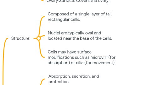
15.2.3.3. Ovary Surface: Covers the ovary.
15.3. Simple Columnar Epithelium
15.3.1. Structure:
15.3.1.1. Composed of a single layer of tall, rectangular cells.
15.3.1.2. Nuclei are typically oval and located near the base of the cells.
15.3.1.3. Cells may have surface modifications such as microvilli (for absorption) or cilia (for movement).
15.3.2. Functions:
15.3.2.1. Absorption, secretion, and protection.
15.3.2.2. Movement of mucus or reproductive cells (in ciliated types).
15.3.3. Locations:
15.3.3.1. Digestive Tract (Stomach to Rectum): Absorptive cells in the intestine are often equipped with microvilli (forming the brush border) to increase surface area for absorption.
15.3.3.2. Gallbladder: Absorbs water and concentrates bile.
15.3.3.3. Uterine Tubes (Fallopian Tubes): Ciliated columnar epithelium moves the ovum towards the uterus.
15.3.3.4. Bronchi: Ciliated columnar epithelium helps in moving mucus and trapped particles out of the lungs.
15.4. Psuedostratified Epithelium
15.4.1. Structure:
15.4.1.1. Appears to be stratified (having multiple layers) because the nuclei are at different levels, but all cells are in contact with the basement membrane, making it technically simple.
15.4.1.2. Typically, cells are columnar, with some reaching the surface and others not.
15.4.1.3. Often has cilia and goblet cells (mucus-secreting).
15.4.2. Functions:
15.4.2.1. Secretion, particularly of mucus.
15.4.2.2. Propulsion of mucus by ciliary action.
15.4.3. Locations:
15.4.3.1. Respiratory Tract (Trachea, Bronchi): Known as respiratory epithelium, where cilia move mucus and trapped particles out of the lungs.
15.4.3.2. Male Reproductive Tract (Epididymis): Non-ciliated variant with stereocilia involved in absorption.