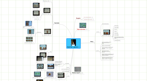
1. Strabismus (squint)
1.1. Non-comitant
1.1.1. the degree of misalignment varies with gaze position
1.1.2. suggests a recent paretic or restrictive aetiology ("paralytic")
1.2. Comitant
1.3. Tropia (a manifest deviation)
1.4. Phoria (a latent deviation)
1.5. Slides
1.5.1. Slide 1
2. Examination
2.1. In different gaze positions
2.1.1. Neuro-ophthalmology illustrated, Bousser et al
2.2. Cover/uncover
2.3. Cross-cover test
2.3.1. 2nd picture
2.3.2. 3rd picture
2.3.3. 4th picture
2.4. Red glass test
2.4.1. Diagnostic use
2.4.1.1. 3rd nerve palsy, 4th nerve in tact?
2.4.1.2. Hypertropia
2.4.1.3. Left Adduction Paresis
2.5. Slides
2.5.1. Examination 1
2.5.2. Examination 2
3. 4th Nerve Palsy
3.1. Neuro Anatomy
3.1.1. Neuro Anatomy
3.2. Clinical Picture
3.2.1. Clinical Picture
3.3. Clinical investigation
3.3.1. Additional investigations
3.4. A possible Differential Diagnosis
3.4.1. Picture
3.4.2. Reasoning
4. Navigation
4.1. EFNS Conference overview
4.2. Free Teaching Course overview
5. History
5.1. Monocular
5.1.1. usually ophthalmological
5.1.2. Differential Diagnosis
5.1.2.1. refractive error
5.1.2.2. corneal disease
5.1.2.3. iris injury
5.1.2.4. cataract
5.1.2.5. media opacity
5.1.2.6. macular disease
5.1.2.7. primary or secondary visual cortex disorder (usually bilateral monocular diplopia
5.1.2.8. Conversion syndrome
5.2. Binocular
5.2.1. Nature of the separation of the two images: horizontal / vertical / oblique
5.2.2. Direction of maximal separation
5.2.3. Onset and progression: sudden onset with improvement / constant / progressive / intermittent
5.2.4. Exacerbating and relieving factors: blinking / worsening at end of day
5.2.5. Associated symptoms: perioribital pain / new headache / intermittend ptosis / visual loss / proximal limb weakness
5.2.6. Past medical and family history: childhood strabismus / childhood glasses / eye occlusion, risk factors -- not complete list -
