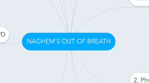
1. • Treatment
1.1. Bronchodilators
1.1.1. • Selective β2 Agonists (SABA & LABA) • Theophylline • Muscarinic Antagonists
1.2. Anti-inflammatory
1.2.1. Corticosteroids Leukotriene modifiers Anti-IgE drugs
1.3. Mast cells stabilizers
1.3.1. Cromolyn Ketotifen
2. • Investigations
2.1. X-RAY
2.2. blood test (IgE AB)
2.3. lung function test
2.3.1. Peak Expiratory Flow Rate Spirometry Bronchprovocation Challenge Test Arterial Blood Gas
3. 4. Hypersensitivity Type I
3.1. B-cells are stimulated (by CD4+TH2 cells) to produce IgE antibodies specific to an antigen. Mast cells and basophils coated by IgE antibodies are "sensitised." Later exposure to the same allergen cross-links the bound IgE on sensitised cells, resulting in degranulation and the secretion of pharmacologically active mediators
4. 5. COPD
4.1. Emphysema
4.1.1. emphysema results from the destructive effect of high protease activity in subject with low antiprotease activity. by stimulation of IL8, LTB4, TNF which attract and activate neutrophils cause release of elastase and tissue damage
4.2. CHRONIC BRONCHITIS
4.2.1. Chronic inflammation
4.3. BRONCHIECTASIS
4.3.1. After bronchial obstruction normal clearing mechanisms are impaired there is pooling of secretions and there is inflammation of the airway also severe infections of the bronchi lead to inflammation
4.4. Pneumothorax
4.4.1. is the presence of air in the pleural cavity . Itmay occur with traumatic perforation of the pleura or may be“spontaneous
5. • Definition
5.1. • Definition
5.1.1. chronic inflammatory disorder of the airways that causes recurrent attacks of breathlessness
5.2. • Signs & Symptoms
5.2.1. Wheeze Chest tightness Cough dyspnoea
5.3. • Risk Factors
5.3.1. Family history. Allergies Smoking Obesity Air Pollution Viral respiratory infections
5.4. • Stages
5.4.1. Step 1 – mild intermittent asthma Step 2 – mild persistent asthma Step 3 – moderate persistent asthma Step 4 – severe persistent asthma
5.5. • Pathophysiology
5.5.1. chronic inflammation of the conducting zone of the airways results in increased contractability of the surrounding smooth muscles leads to bouts of narrowing of the airway and the classic symptoms of wheezing
5.6. • Complication
5.6.1. Fatigue psychological problems pneumonia collapse respiratory failure status asthmaticus
6. Mechanical defenses
7. Brain initiates inspiratory command The inspiratory muscles contract Intrapleural pressure becomes more negative The transpulmonary pressure increases Alveoli expand (according to Boyle’s law Alveolar pressure falls below atmospheric pressure Air flows into the alveoli from the atmosphere through airways
8. Histology of Respiratory System
8.1. Trachea
8.1.1. Mucosa Submucosa Cartilage and Smooth Muscle Layer Adventitia
8.2. Bronchi
8.2.1. mucosa with isolated plates of hyaline cartilage Small mucous and serous glands The lamina propria
8.3. Bronchioles
8.3.1. they lack both mucosal glands and cartilage the epithelium is still ciliated pseudostratified columnar Clara cells
8.4. Respiratory Bronchioles
8.4.1. Smooth muscle and elastic connective tissue
8.5. alveolar sacs
8.5.1. network of elastic and reticular fibers network of capillaries
8.6. Alveoli
8.6.1. Between neighboring alveoli lie thin interalveolar septa
9. 2. Physiology of Breathing
9.1. inspiration
9.2. expiration
9.2.1. Brain ceases inspiratory command • Inspiratory muscles relax. Thoracic volume decreases, causing intrapleural pressure to become less negative Decreased transpulmonary pressure allows the increased alveolar elastic recoil Decreased alveolar volume increases alveolar pressure above atmospheric pressure thus establishing a pressure gradient for airflow
10. 3. Immunity
10.1. Innate immunity
10.1.1. soluble mediators
10.1.2. Phagocytic cells like macrophages and dendritic cells
10.2. Adaptive immunity
10.2.1. Humeral Immunity (B Cell Plasma Cell Ab)
10.2.2. Cellular Immunity (TH1 + TH2 + Cytotoxic Cells + APC)
10.2.3. In RS it includes:
10.2.3.1. epithelial compartment MALT lymph nodes

