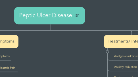
1. Etiology/ Risk Factors
1.1. Non Infectious Causes
1.1.1. Genetic predisposition to ulcer formation
1.1.2. Poor cell restitution
1.1.3. Excessive acid secretion
1.1.4. Stress
1.1.5. Copious amounts of alcohol intake
1.1.6. Smoking
1.1.7. Ingestion of aspirin and NSAIDS (non-steroidal anti-inflammatory drugs)
1.1.8. Chronic use of drugs
1.1.8.1. Steroids
1.1.8.2. Potassium or iodide compounds
1.2. Infectious Causes
1.2.1. Bacteria
1.2.1.1. Helicobacter pylori bacterium
1.2.1.2. Increased likelihood of infection if there is a polymorphism in IFNGR1 (interferon gamma receptor-1)
1.3. Variation in Lewis Blood group antigen
1.3.1. Epithelial receptor for H. Pylori
1.3.2. Does not have the correct antigen to get rid of the infection/ microorganisms
1.3.3. A gene on chromosome 11p15.5 that encodes a glycoprotein of gastric and respiratory tract epithelia, protecting mucosa from infection and chemical damage by binding to microorganisms and particles
2. Incidence/ Prevalence
2.1. Duodenal Ulcers
2.1.1. Common in both men and women
2.1.2. Occurs most often in 40-50 year olds, though with some incidence in young adults with in stressful situations
2.1.3. Occurs in 10% of the US population
2.1.3.1. High risk of recurrence
2.2. Gastric Ulcers
2.2.1. Aged 60-70 years and more prevalent in men
2.2.2. Perforation of ulcer has three times greater of a risk of death than duodenal Ulcers
2.2.2.1. Due to age of patient
2.3. People who are at a greater risk
2.3.1. African Americans
2.3.1.1. Asians/ Pacific Islanders
3. Diagnostics
3.1. Barium radiographic studies
3.1.1. Presence of ulcers often as a protrusion on radiographic examination
3.1.2. Barium study highlights presence of ulcer in stomach or duodenum
3.2. Esophagogastroduodenoscopy
3.2.1. Presence of mucosal ulcerations in the stomach or duodenum
3.2.2. Flexible endoscopy to allow visualization of mucosa
3.3. Serum gastrin levels tested
3.4. Hemoglobin/ Hematocrit test
3.5. Complete blood count
3.6. Tests for H. Pylori
3.6.1. Antibody detection
3.6.2. Urea breath test
3.6.2.1. Identifying enzymatic presence and use of bacterial urease
3.6.2.2. Serum, plasma, urine, whole blood
3.6.3. fecal antigen testing detects presence of H. pylori antigens in stools
4. Signs/ Symptoms
4.1. Major symptoms
4.1.1. Epigastric Pain
4.1.1.1. Sharp, burning, or gnawing sensations occur
4.1.1.2. Can be constant or have no pattern whatsoever
4.1.2. Weight loss/ Anorexia
4.1.2.1. Due to pain that occurs when ingesting foods and liquids
4.2. Signs
4.2.1. Pale mucous membranes/ skin
4.2.1.1. Anemia
4.2.1.1.1. Acute or chronic blood loss
4.2.2. Black/ tarry stool
4.2.2.1. Only if massive bleeding is occurring should there be red or bloody stool
4.2.3. Bowel sounds hyperactive
4.2.4. Palpating shows epigastric tenderness
5. Treatments/ Interventions
5.1. Analgesic administration
5.2. Anxiety reduction
5.3. Environmental management
5.4. Engaging in comforting actions
5.5. Pain management
5.6. Medication management
5.7. Patient controlled analgesia assistance
5.8. H2 antagonist
5.8.1. Reduces acid secretion to optimize ulcer healing
5.8.2. Cimetidine, rantidine, famotidine, nizatidine
5.9. Proton pump inhibitors
5.9.1. Optimizes ulcer healing by binding to the proton pump of parietal cell and inhibiting secretion of hydrogen ions into gastric lumen
5.9.2. Omeprazole, esomeprazole, lansoprazole, rabeprazole, pantoprazole
6. Evidence Based practice health policy
6.1. Discontinuation of low-dose aspirin therapy is often recommended to reduce bleeding risks associated with peptic ulcer disease. However, investigators suggest that discontinuation of aspirin therapy may increase the risk of cardiovascular-related mortality.
6.2. In a retrospective study among 118 patients treated for peptic ulcer bleeding who were also previously taking low-dose aspirin, aspirin therapy was discontinued in 40%. During the first 6 months of follow-up among patients with cardiovascular comorbidities, 31% of patients who discontinued aspirin therapy experienced an acute cardiovascular event or death compared to 8% of patients who continued aspirin therapy.
6.3. Patients who discontinued aspirin therapy were 6.3 times more likely to experience an acute cardiovascular event or death within 6 months compared to patients who continued aspirin therapy.
7. Associated Diseases
7.1. Type B Chronic Gastritis
7.1.1. Helicobacter pylori bacterium also a cause of this disease
7.2. Hyperparathyroidism
7.3. Chronic Lung Disease
7.4. Alcoholic Cirrhosis
7.5. Zollinger-Ellison Syndrome
7.5.1. Stomach produces too much acid, therefore ulcers form
7.5.1.1. Resulting in diarrhea and stomach pain
