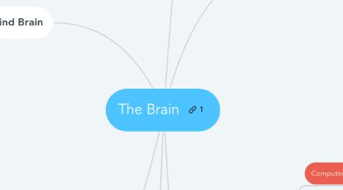
1. Hind Brain
1.1. Cerebellum
1.1.1. Responsible for a number of functions including motor skills such as balance, coordination, and posture.
1.2. Medulla
1.2.1. The medulla oblongata helps regulate breathing, heart and blood vessel function, digestion, sneezing, and swallowing. This part of the brain is a center for respiration and circulation. Sensory and motor neurons (nerve cells) from the forebrain and midbrain travel through the medulla.
1.3. Pons
1.3.1. It bridges sensory information between the left and right hemispheres of the brain.
1.4. Reticular Formation
1.4.1. Its functions can be classified into 4 categories: motor control, sensory control, visceral control, and control of consciousness.
2. Language
2.1. Broca's Area
2.1.1. controls the ability to produce language
2.2. Wernicke's Area
2.2.1. responsible for the comprehension of speech.
3. Special Topics
3.1. Split Brain
3.1.1. when the corpus callosum connecting the two hemispheres of the brain is severed to some degree.
3.2. 1 hemisphere
3.2.1. A situation where one hemisphere is missing from birth, causing impairment of certain functions but also allowing the brain to develop radically through its plasticity
3.3. Concussions
3.3.1. A mild form of traumatic brain injury, in which there is a traumatically induced alteration in mental status, with or without an associated loss of consciousness.
4. Emotional Brain
4.1. Limbic System
4.1.1. Thalamus
4.1.1.1. It works to correlate several important processes, including consciousness, sleep, and sensory interpretation.
4.1.2. Amygdala
4.1.2.1. It is a section of the brain that is responsible for detecting fear and preparing for emergency events.
4.1.3. Hippocampus
4.1.3.1. It plays a very important role in storing our memories and connecting them to our emotions, among other roles.
4.1.4. Cingulate Cortex
4.1.4.1. It receives inputs from the thalamus and the neocortex, and projects to the entorhinal cortex via the cingulum.
4.1.5. Hypothalamus
4.1.5.1. It controls the pituitary gland and also regulates homeostasis (hunger, thirst, body temperature, and the like).
5. The Cortex
5.1. Left/Right Hemisphere
5.1.1. Cerebral hemispheres that control functions on the opposite side on the body.
5.2. Corpus Callosum
5.2.1. it is responsible for allowing the two hemispheres to communicate with each other and share information.
5.3. Lobes
5.3.1. Parietal
5.3.1.1. It functions in processing sensory information from the various parts of the body.
5.3.2. Temporal
5.3.2.1. It is involved in vision, memory, sensory input, language, emotion, and comprehension.
5.3.3. Frontal
5.3.3.1. The frontal lobes are important for controlling thoughts, reasoning, and behaviors.
5.3.4. Occipital
5.3.4.1. Its primary function is the processing of vision and visual information.
5.4. Motor Cortex
5.4.1. The motor cortex region is responsible for all voluntary muscle movements.
5.5. Somatosensory Cortex
5.5.1. This part of the brain processes sensations, or external stimuli, from our environment. The somatosensory cortex receives all sensory input from the body.
6. Brain Scans
6.1. Computed Tomography
6.1.1. A radiographic method for swiftly generating complex, 3D visuals of the mind or other soft tissues.
6.2. Magnetic Resonance Imaging
6.2.1. used for studying the functions of the brain (or any living tissue) without surgery. Images are obtained by using a strong magnetic field.
6.3. Electroencephalogram
6.3.1. An electroencephalogram (EEG) is a recording of the electrical waves of activity that occur in the brain, and across its surface. Electrodes are placed on different areas of a person's scalp, filled with a conductive gel, and then plugged into a recording device. The brain waves are then attracted by the electrodes, travel to the recording device and then amplified so that they can be more easily seen and examined.
6.4. Positron Emission Tomography
6.4.1. a scanning method that enables psychologists and doctors to study the brain (or any other living tissue) without surgery. PET scans use radioactive glucose (instead of a strong magnetic field) to help study activity and locate structures in the body.
6.5. Functional MRI
6.5.1. a brain imaging technique that detects magnetic changes in the brain's blood flow patterns. This technique is a combination of Magnetic Resonance Imaging (MRI) and Positron Emission Tomography (PET) scans and is useful for detecting changes in activation of different centers of the brain.

