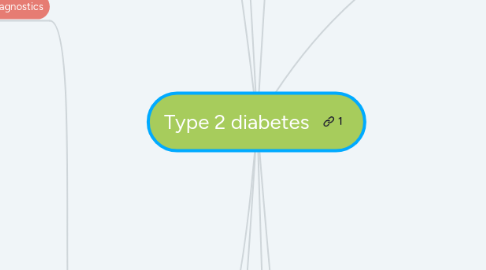
1. Time/course
1.1. Progression
1.1.1. Earliest signs of pre diabetes is insulin resistance in the patient. At this stage however the pancreatic B-cell is still able to compensate by secreting insulin. This is called the pre-diabetes stage.
1.1.2. From here patients progress to the impaired glucose tolerance stage of diabetes. This is caused by hyperinsulinemia. Over time the B cells become refractory to glucose causing an insulin deficiency to develope (Fonseca, 2009).
1.1.3. Later in the diseases progression the B cell will deteriorate even further leading to an absolute insulin deficiency. This will result in the B cell becoming unresponsive to medical interventions.
1.1.4. At this point the patient requires exogenous insulin. This is the insulin-requiring stage of type 2 diabetes.
1.2. Complications
1.2.1. Diabetic foot
1.2.2. Cardiovascular
1.2.2.1. High blood pressure
1.2.2.2. Stroke
1.2.2.3. Atherosclerosis
1.2.3. Kidney
1.2.3.1. Nephropathy
1.2.4. Skin
1.2.4.1. Skin infections
1.2.5. Opthalmic
1.2.5.1. Cataracts
1.2.5.2. Glaucoma
1.2.5.3. Retinopathy
1.2.6. Emergency
1.2.6.1. Diabetic ketoacidosis
1.2.6.2. Myocardial infarction
1.2.7. Nerve
1.2.7.1. Neuropathy
1.2.8. Blood vessels
1.2.8.1. Microangiopathy
1.2.8.2. Microvascular complications
1.2.8.3. Peripheral vascular disease
1.2.8.4. Ulceration
2. Signs and symptoms
2.1. Type 2 diabetes
2.1.1. Fatigue
2.1.2. Blurred vision
2.1.3. Unintended weight loss
2.1.4. Increased hunger
2.1.5. Frequent urination
2.1.6. Increased thirst
2.1.7. Slow healing sores
2.1.8. Frequent infections
2.1.9. Numbness and tingling in the hands and feet
2.1.10. Areas of darkened skin
2.2. Diabetes ketoacidosis
2.2.1. Vomiting
2.2.2. Abdominal pain
2.2.3. Nausea
2.2.4. Sweet smelling breath
2.2.5. Shortness of breath (SOB)
3. Diagnostics
3.1. Conventional diagnostics
3.1.1. Laboratory tests
3.1.1.1. HbA1c (glycated haemoglobin test)
3.1.1.1.1. Glycated haemoglobin test HbA1c: It indicates blood sugar levels from the past 2-3 months. Levels above 6.5% on two seperate tests is indicative of diabetes.
3.1.1.2. Random blood glucose test
3.1.1.2.1. If the A1c test is not conisisten the following tests may be used. - random blood glucose test: higher than 11.11 mmol/L is indicative of diabetes("Type 2 diabetes - Diagnosis and treatment Mayo Clinic", 2021).
3.1.1.3. Fasting blood glucose test
3.1.1.3.1. 7mmol/l or higher is considered diabetes.
3.1.1.4. Oral glucose tolerance test
3.1.1.4.1. Fasting (8 hours), 75g of glucose is then administered, blood is then collected from sample and two hours post glucose uptake. Considered diabetes if after 2 hours if blood glucose is equal or more than 200mg/dL
3.2. Osteopathic diagnostics
3.2.1. Case history
3.2.1.1. Insidious onset.
3.2.1.2. Primary complaint is fatigue.
3.2.1.3. Previous history of coronary artery disease, hypertension, claudication, polydipsia, nocturia, weight loss and fatigue.
3.2.1.4. Has the patient had a cerebrovascular accident ?
3.2.1.5. History of neuropathy or symptoms of peripheral neuropathy
3.2.1.6. Evaulate their diet and exercsise levels.
3.2.1.7. Do they have any foot ulcers ?
3.2.2. Does the patient have hyperlipedemia ? When was their last test ?
3.2.3. Physical exam
3.2.3.1. Systems examination
3.2.3.1.1. Neurological examination of lower limb.
3.2.3.1.2. Observe for skin changes (coolness,paleness, dryness, pimples and abnormal pigmentation).
3.2.3.1.3. Opthalmic examination assessing the retina, macula and optic disc.
3.2.3.1.4. Vital signs examination assessing blood pressure, temperature and weight.
3.2.3.2. Palpation
3.2.3.2.1. Assess for tissue changes such as doughy, ropey and thickened interstitial tissue.
3.2.3.2.2. Assess for viscerosomatic reflex changes at the T11-L2 area(Licciardone et al., 2007).
3.2.3.3. Foot examination
3.2.3.3.1. Assess the dorsalis pedis and posterior tibailis pulses.
3.2.3.3.2. Observe for skin changes on the foot such as ulcerations.
3.2.3.4. Observation
3.2.3.4.1. Observe physical size (waist height ratio)
4. Treatment and management
4.1. Conventional
4.1.1. Treatment
4.1.1.1. Medications
4.1.1.1.1. Metformin
4.1.1.1.2. Sulfonulyrias
4.1.1.1.3. Glinides
4.1.1.1.4. Insulin therapy
4.1.1.2. Weight loss surgery
4.1.1.3. Alternative medications
4.1.1.3.1. Magnesium
4.1.1.3.2. Cinnamon
4.1.1.3.3. Chromium
4.1.2. Managment
4.1.2.1. Lifestyle
4.1.2.1.1. Diet changes
4.1.2.1.2. Inreased physical activity
4.1.2.1.3. Weight loss
4.1.2.1.4. Healthy lifestyle education
4.2. Osteopathic
4.2.1. Treatment
4.2.1.1. Rib raising at 2nd and 4th rib
4.2.1.2. Myofascial release of hypertonic tissue
4.2.1.3. Kneading of hypertonic tissue
4.2.2. Managment
4.2.2.1. Education
4.2.2.2. Lifestlye modifications
4.2.2.2.1. Increased physical activity
4.2.2.2.2. Dietary changes
4.2.2.2.3. Weight loss
5. Pathophysiology
5.1. Type 2 diabtes is caused by a combination of two primary factors.
5.2. 1. Defective insuline secretion by the B-cells of the pancreas.
5.3. 2. Tissue inability to respond appropraitely to the insulin (Mukhamedzhanov & Esyrev, 2013). These two factors are caused by the following mechanisms.
5.4. Malfunctioning of the feedback loop between the secretion of insulin and it's function causes high levels of glucose in the blood.
5.5. As the disease progresses Beta cells begin to change resulting in B-cell dysfunction. This results in reduced insulin secretion which then limits the bodys ability to maintain glucose levels (Galicia-Garcia et al., 2020).
5.6. Simultaneously insulin resistance will increase glucose production in the liver, however will decrease the glucose uptake in the muscle and adipose tissue.
5.7. When both B-cell dysfunction and insulin resistance are present the patients body will become hyperglycaemic leading to the progression of type 2 diabetes.
6. Epidemiology
6.1. Co-morbidities
6.1.1. Obesity
6.1.2. Dyslipedemia
6.1.3. Depression
6.1.4. Retinopathy
6.1.5. Cardiovascular
6.1.5.1. Hypertension
6.1.5.2. Coronary heart disease
6.1.5.3. Peripheral vascular disease
6.1.6. Kidney
6.1.6.1. Chronic kidney disease
6.1.6.2. Renal disease
6.2. Obesity
6.3. Prevalence
6.3.1. Average patient has had diabetes for 4-7 years before diagnosis.
6.3.2. 1.2 million Australians have diabetes.
6.3.3. 6.1% of men and 4.6% of women suffer from diabetes.
6.3.4. It is twice as high for people living in low socioeconomic areas.
6.3.5. 280 Australians develope diabetes everyday.
6.4. Risk factors
6.4.1. Lifestyle risk factors
6.4.1.1. Cigarette smoking
6.4.1.2. Overweight
6.4.1.3. High blood pressure
6.4.1.4. Sedentary lifestyle
6.4.1.5. Unhealthy eating habits
6.4.1.6. Hypercholesterolaemia
6.4.2. Genetic/biological risk factors
6.4.2.1. Family history of diabetes
6.4.2.2. Gene mutations associated with diabetes
6.4.2.3. Close family member with diabetes
6.4.2.4. Age over 45
6.4.3. Environmental risk factors
6.4.3.1. Area of low walkability
6.4.3.2. Lack of green spaces in neighbourhood
6.4.3.3. Air pollution
6.4.4. Race/ethnicity risk factors
6.4.4.1. African American
6.4.4.2. Asian American
6.4.4.3. Aboriginal and Torrest strait Islander
6.4.4.4. Pacific islander
7. Osteoarthirits
7.1. Conventional
7.1.1. Treatment
7.1.1.1. Medications
7.1.1.1.1. NSAIDS
7.1.1.1.2. Paracetamol
7.1.1.1.3. Topical therapies
7.1.1.1.4. Intra-articular corticosteroid injections
7.1.1.1.5. Opoids
7.1.1.1.6. Duloexetine
7.1.1.1.7. Complementary medications (cod liver oil, glucosamine sulphate)
7.1.1.2. Joint replacement surgery
7.1.2. Management
7.2. Osteopathic
7.2.1. Treatment
7.2.1.1. Accupuncture
7.2.1.2. Osteopathic manipulation of knee joint and peripheral joints.
7.2.1.3. Soft tissue massage of adjacent tissues.
7.2.2. Managment
7.2.2.1. Educational advice
7.2.2.1.1. Education on disease process, complications, weight managment and diet.
7.2.2.2. Lifestyle changes
7.2.2.2.1. Diet plans
7.2.2.2.2. Physical activity prescription
7.2.2.2.3. Weight loss management
8. Examinations for OA
8.1. Systems
8.1.1. Cardiovascular: Anxiety attack
8.1.2. Respiratory: SOB
8.1.3. Gastrointestinal: Type 2 Diabetes
8.1.4. Mental health: Had 1 anxiety attack
8.2. Physical tests
8.2.1. Standing
8.2.1.1. Observation: uneven weight bearing, obese
8.2.2. Seated
8.2.2.1. Lower Limb neuro: altered senses around medial knee, Myotomes +5, Reflexes +2
8.2.3. Supine
8.2.3.1. Ruling in
8.2.3.1.1. Extensor Quad lag +ve
8.2.3.1.2. Patella Grind +ve
8.2.3.1.3. Muscle wasting
8.2.3.1.4. Palpation: Tenderness around knee joint
8.2.3.1.5. Knee AROM: Reduced extension, pain on end-ROM
8.2.3.2. Ruling out
8.2.3.2.1. McMurrays -ve
8.2.3.2.2. Valgus/Varus -ve
8.2.3.2.3. Posterior drawer -ve
8.2.3.2.4. Anterior Drawer -ve
8.2.4. Side lying
8.2.4.1. Obers test: -ve
8.2.4.2. Palpation NAD, pain on knee when stacked together
8.2.5. Prone
8.2.5.1. Palpation: NAD
