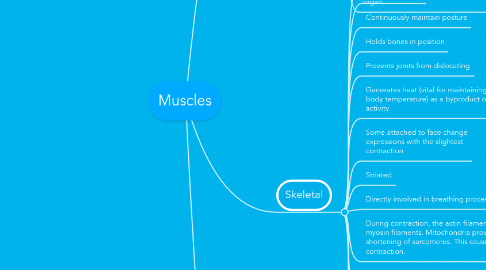
Muscles
par Hannah K


1. Smooth
1.1. Found in walls of hollow organs (intestines, bladder, stomach, uterus, etc.)
1.2. Involuntary
1.2.1. Involved in 'housekeeping' of the body
1.3. Involved in 'housekeeping' functions of the body
1.4. Lack of visible striations
1.5. Made up of thin-elongated muscle cells, fibres
1.6. Interlace to form sheets or layers of muscle tissue
1.7. Controls slow, involuntary movements (contraction of smooth muscle tissue in walls of intestine and uterus)
1.8. Single nucleus
1.9. Lacks transverse tubules
1.10. Contracts and relaxes slowly
1.11. Rhythmic
1.12. Self-exciting
1.13. Cells are elongated with tapering ends
1.14. Contain actin and myosin in myofibrils that extend the length of the cells
1.15. Sarcoplasmic reticulum is not well developed
1.16. Multiunit muscle fibers are separate, contracts only in response to stimulation by motor nerve impulses or certain hormones, and are found in the irises of the eyes and walls of blood vessels
1.17. Visceral muscle is composed of sheets of spindle-shaped cells in close contact of one another, found in walls of hollow organs, fibers can transmit impulses from cell to cell (Self-exciting), are rhythmic, and are responsible for the wavelike motion (peristalsis) that occurs in certain tubular organs and helps to force the contents along the length of the organ
2. Skeletal
2.1. Cover skeleton and give it shape
2.1.1. G
2.2. Voluntary control
2.3. Continuously maintain posture
2.4. Holds bones in position
2.5. Prevents joints from dislocating
2.6. Generates heat (vital for maintaining normal body temperature) as a byproduct of muscle activity
2.7. Some attached to face change expressions with the slightest contraction
2.8. Striated
2.9. Directly involved in breathing process
2.9.1. Dur
