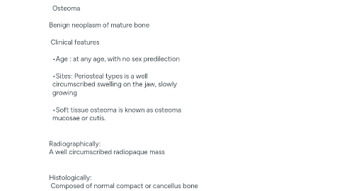
1. Classification of bone lesions • Hereditary bone diseases: Osteogenesis imperfect, Osteopetrosis, Cherubism, Cleidocranial dysplasia,& Achondroplasia. • Inflammatory bone diseases: Periapical granuloma, cyst, and abscess in addition to dry socket and osteomyelitis. • Hormonal disease: Hyperparthyroidism. • Nutritional deficiency diseases: Rickets, Scurvy and Osteomalacia • Giant cell lesions (Multinucleated giant cell lesions) Central giant cell granuloma, Fibrous dysplasia, Aneurismal bone cyst, Paget's disease of bone. Bone tumors (neoplasm) A: Non-odontogenic neoplasm B: Odontogenic neoplasm Non-odontogenic neoplasm A: Benign neoplasm As osteoma, osteoid osteoma, Chondroma osteochondroma, & osteoblastoma B: Malignant neoplasm Primary bone tumors Secondary tumors Osteoma Benign neoplasm of mature bone Clinical features •Age : at any age, with no sex predilection •Sites: Periosteal types is a well circumscribed swelling on the jaw, slowly growing •Soft tissue osteoma is known as osteoma mucosae or cutis. Radiographically: A well circumscribed radiopaque mass Histologically: Composed of normal compact or cancellus bone Treatment : Surgical removal Osteogenic sarcoma It is a tumor of malignant osteoblasts Clinical features: •Occurs in all bones of the skeleton. Age: the second and third decades The chief signs and symptoms are swelling and pain at the affected site, growth of the tumor is rapid and it is estimated that 6% of osteosarcomas arise in the jaws, the mandible is more often involved than the maxilla (2 to I ratio). The most common symptoms of jaw tumors swelling, pain, lose teeth, paresthesia, toothache, bleeding, and headache. Radiographic features: Sun ray (sunburst) pattern Treatment surgical resection followed by extensive chemotherapy Multiple myeloma A malignant neoplasm of plasma cells. •Tumor cells secrete large quantities of immunoglobulin that are detected in the serum by electrophoresis. •Any of the five classes of antibodies may be produced but lgG is the most common. When excreted in the urine where they are referred to as Bence Jones proteins. Clinical features and characteristics: • Bone pain caused by multiple plasma cell tumors within bone marrow is often the earliest symptom. • Normocytic, normochromic anemia. • Hypercalcemia develops as bone is resorbed. • 10% of myeloma patients develop amyloidosis caused by the precipitation of immunolgobulins within organs and tissues. • Pathologic fracture of involved bones is a feature of late stage disease. Radiographic features: Multiple "punched-out" radiolucent lesions in bone are the characteristic feature Histologic Features: Group of plasma cells. Treatment: remission may be with systemic chemotherapy using prednisone & cyclophosphamide. Fibrous dysplasia The basic defect appears to be a benign proliferation of fibroblasts arising within bone marrow. Classification: (monostotic, polyostotic & familial or hereditary). A: monostotic types a single bone is involved, and not progressed to polyostotic type. •Clinically:- Common in maxilla than mandible-mainly labial or buccal plate of bone From 4-18 years of age • Facial deformity • Mal -alignment of teeth • Bone affected: skull, clavicle, pelvis, scapula & long bones. Etiology: Unknown, it may be trauma or local infection • Affected bone become enlarged with cortical thinning. • pathologic fractures Radiographic features: are so variable, purely radiolucent forms or a finely trabeculated radiodensity, the so called "ground glass" appearance (most common). Ground glass appearance B: Polyostotic fibrous dysplasia In addition to multiple bone involvement, there are: • Large patches of melanotic skin pigmentation referred to as the "Jaffe" variant (cafe-au-lait spots). • Bone lesions with skin pigmentation and endocrinopathy are referred to as "Albright's syndrome.“ Treatment surgical curettage followed by bone graft Cherubism •It is inherited disease, characterized by marked fullness of the face. •Involvement of the maxilla causes expansion with upward displacement of the eyes producing the so- called "heavenly gaze“ •The name "cherubism" comes from the cherubic facial appearance . •Etiology: mutations in the SH3BP2 gene on chromosome #4 •age 4-8 years. •Males affected more than females (2:1) Radiographic features: appears as symmetrical bilateral multilocular radiolucent lesions. Typically, lesions begin in the mandibular rami and advance anterior Treatment: • The disease may be self-limiting, and may stop growing and regress during the teens. • Surgical curettage may achieve cosmetic improvement in those patients with unsightly jaw enlargement. Paget's disease of bone (Osteitis deformans) • Paget disease is a skeletal disease of unknown cause. • Recent evidence indicates this disease is triggered by infection of osteoclasts with the measles virus or respiratory syncytial l virus (RSV). • It may affect a single bone (monostotic form) or multiple bones (polyostotic form). • In the early years, osteoclastic resorption of normal bone is accelerated. Clinical features: ❑ It is uncommon for Paget disease to occur before age 40. ❑ Men and women are affected equally. ❑ crippling, bowing deformities. ❑ If the base of the skull is affected, narrowing of cranial foramina may cause deafness and blindness. • The maxilla is more often involved than the mandible. • Involved bones become progressively larger. • Hats and dentures may no longer fit and in medical lore, these signs have become classic. • When jaws are involved, the teeth often show hypercementosis • Teeth may drift apart as the jaws enlarge. • Patients with Paget disease have an increased risk of developing sarcoma of bone. Radiographic features: The radiographic appearance varies with the stage of the disease. • Early stage: osteolysis dominates and the lesion is radiolucent. • Middle stage: apposition of "paget bone" creates islands of density within the radiolucent lesions. these islands lack the normal trabecular pattern and are homogeneously dense. because they resemble tufts of cotton, they are called "cotton wool" densities. • late stage: osteoblastic apposition of paget bone continues as osteoclastic activity subsides. the bone becomes homogeneously dense. Histological features: Osteoclastic resorption and osteoblastic apposition, sometimes in the same field. ❑ "Mosaic" trabeculae. ❑ Fibrous connective tissue replacement of the normal fatty marrow. ❑ Vascular dilation. ❑ Lymphocytic infiltration suggesting an inflammatory basis for the disease osteopetrosis It is a rare inherited disease of bone in which there is failure of normal osteoclastic resorption. Although osteoclasts fail to resorb bone, osteoblasts exhibit normal function. The imbalance between osteoblasic of bone and osteoclastic resorption leads to increasing bone density. May be due to defect in carbonic anhydrase enzyme. Clinical features: • osteopetrosis may be leading to blindness, facial paralysis, and deafness, due to the increased pressure put on the nerves by the extra bone. • Enamel hypoplasia, delay tooth eruption and difficult in tooth extraction. Radiographic features: Achondroplasia • is a form of short-limbed dwarfism. • in achondroplasia the problem is not in forming cartilage but in converting it to bone (a process called ossification), particularly in the long bones of the arms and legs. • Teeth are larger than the jaws • Sometimes patient are prone to gingivitis due to mouth breathing