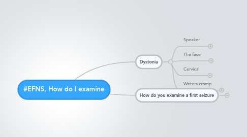
1. Dystonia
1.1. Speaker
1.1.1. Marie Vidailhet Paris, France
1.1.2. Due to copyright issues / patient privacy no images are included, but this session has many nice images / videos
1.2. The face
1.2.1. Only partly reported
1.2.2. Not dystonia
1.2.2.1. hemimasticatory spasm in hemifacial atrophy, Parry Romberg Syndrome
1.2.2.2. permanent, tonic, unilateral
1.2.3. Odd or complex dystonia
1.2.3.1. Secondary causes of dystonia may be suspected
1.2.3.2. Eyelid apraxia w PSP
1.2.3.3. STN stimulation parkinson
1.2.3.4. Involvement of lower face
1.2.3.4.1. neuro- acanthocytosis
1.2.3.4.2. PANK2
1.2.3.4.3. Neuroferritinopathies
1.2.3.4.4. Wilson's Disease
1.2.4. Practical consequences
1.2.4.1. If primary dystonia
1.2.4.1.1. Botulinum toxin injections
1.2.4.2. In very severe cases
1.2.4.2.1. DBS
1.3. Cervical
1.3.1. classical
1.3.1.1. young adult
1.3.1.2. more frequent in women
1.3.1.3. Symptoms
1.3.1.3.1. torticollis
1.3.1.3.2. retrocollis
1.3.1.3.3. laterocollis
1.3.1.3.4. myoclonic cervical dystonia
1.3.2. Dynamic approach
1.3.2.1. Geste antagoniste
1.3.2.2. Wrinkles
1.3.2.2.1. on the skin may be the signature of muscle contraction and abnormal postures
1.3.2.3. Muscle hypertrophy
1.3.2.4. Pain (muscle tenderness or tension)
1.3.2.5. Predominant abnormal posture (splenius, SCM)
1.3.2.6. role of trunk (posture, gait) abnormal trunk posture
1.3.2.7. etc.
1.3.3. Consierd theneck mobility and posture!
1.3.4. Treatment
1.3.4.1. physical therapy
1.3.4.2. Pain treatment
1.3.4.3. Botulinum toxin
1.3.4.4. very very severe cases DBS
1.3.5. Secondary causes
1.3.5.1. Stiff neck
1.3.5.2. Family history
1.3.5.3. congenital torticollis
1.3.5.3.1. since infancy
1.3.5.4. Post traumatic
1.3.5.4.1. spinal cord lesion
1.3.5.5. Stiff person
1.3.5.5.1. rapid onset, pain, anti GAD Ab
1.3.5.6. Neuroleptics
1.3.5.6.1. Retrocollis
1.3.5.7. Psychogenic
1.3.6. Amplitude limitation and thin sterno cleido mastoid (fibrosis)
1.3.6.1. no botulinum toxin...as there's no muscle
1.3.6.2. surgeon
1.4. Writers cramp
1.4.1. examination
1.4.1.1. write spontaneously: velocity, stop between words, legible or non legible, tremor, presss excessively on the pen
1.4.1.2. abnormal posture; jumping pen
1.4.1.3. write with the non dominant hand: dystonia appears on the dominant hand
1.4.1.4. drawings
1.4.2. Risk of writer's cramp
1.4.2.1. increasesw time spent writing each day
1.4.2.2. reference:Roze e, et al, Brain 2009
1.4.3. Pitfall: compensatory posture hides the real pattern of dystonia
1.4.4. Compensation and dystonia
1.4.4.1. when present the "mirror" phenomenon is most helpful to observe the core of dystonia
1.4.5. Abnormalposture is not always pathological
1.4.5.1. e.g. in left handed writing
1.4.6. Sensory disturbances
1.4.6.1. movement appears when eyes are closed
1.4.6.2. looks like dystonia
1.4.6.3. it's something else...
1.4.6.4. clues
1.4.6.4.1. myokimia that can be seen in CIDP
1.4.6.4.2. lesion in Cervical myelum
1.4.6.4.3. myasthenia gravis can exceptionally mimic normal posture when playing piano (intensive)
2. How do you examine a first seizure
2.1. Author
2.1.1. Paul Boon Ghent, Belgium
2.2. Prevalence
2.3. epilepsy
2.3.1. disease of CNS
2.3.2. disease originating from the brain cortex
2.3.3. characterized by occurence of recurrent seizurs = epilepsy
2.3.4. epileptic seizures
2.3.4.1. definition
2.4. seizure
2.4.1. Classification
2.4.1.1. Focal
2.4.1.2. Partial
2.4.1.3. Generalized
2.5. Underlying cause
2.6. First seizure
2.6.1. 99% self-limiting
2.6.2. general practitioner
2.6.3. emergency room
2.6.4. neurologist
2.6.5. small % not self-limiting --> status epilepticus --> emergency room --> neurologist --> intensive care unit
2.7. OBSERVATION
2.7.1. is of incredible importance
2.7.2. if not observation: clinical history
2.8. Was it a seizure?
2.8.1. Differential diagnosis
2.8.1.1. Syncope
2.8.1.2. cardiac arrhytmias
2.8.1.3. hypoglycaemia
2.8.1.4. carotid sinus hypersensitivity
2.8.1.5. panic attacks
2.8.1.6. hyperventilation
2.8.2. a detailed account from patient
2.8.2.1. witness or proxy
2.8.2.2. Account on how long the seizure lasted is almost always over estimated by patients/family on a first seizure
2.8.2.3. Was it really the first seizure?
2.8.2.3.1. inrecognised seizures before?
2.8.3. Prodromal symptoms
2.8.3.1. such as typical auras
2.8.3.1.1. epigastricsensations
2.8.3.1.2. presyncopal symptomas such as light-headedness
2.8.3.1.3. unsteady gait
2.8.3.1.4. visual disturbances
2.8.4. Tongue biting
2.8.4.1. not pathognomonic
2.8.4.2. lateral tongue biting is more specific
2.8.5. Incontinence
2.8.5.1. may happen w syncope as well
2.8.6. History of stroke
2.8.7. head trauma
2.8.8. family history of epilepsy
2.8.9. provoking factors
2.8.9.1. alcohol intake or withdrawal
2.8.9.2. drug abuse
2.8.9.3. sleep deprivation
2.8.9.4. exposure to stroboscopic light
2.9. tests
2.9.1. 2.4-8% of patients have a metabolic underlying cause
2.9.2. Imaging
2.9.2.1. CT cerebrum
2.9.2.1.1. 6-10% of CT are abnormal
2.9.2.1.2. 41% of adults have an abnormal CT following first generalisde seizure
2.9.2.1.3. Neuroimaging is necessary after first seizure
2.9.2.1.4. Acute CT scan
2.9.2.2. MRI cerebrum
2.9.2.2.1. MRI preferred on CT
2.9.2.3. Yield increases with age
2.9.2.4. cortical atrophy, cerebral infarction
2.9.2.5. confirming / excluding diagnosis
2.9.3. EEG
2.9.3.1. Necessary in all young patients presenting w a generalised seizure
2.9.3.2. May support diagnosis in older patients
2.9.3.2.1. EEG cannot exclude epilepsy
2.9.4. LAB
2.9.4.1. Alcohol abuse
2.9.4.2. When suspected drug abuse: substance markers
2.9.5. ECG
2.9.6. Lumbar puncture
2.9.6.1. any suspection on CNS infection
2.9.6.2. CT prior to LP is often wise
2.10. Essential diagnositc procedures
2.10.1. clinical examination
2.10.2. assessment of seizure semiology
2.10.3. routine lab tests
2.10.4. LP if necessary
2.10.5. early EEG within 24 hours
2.10.6. MRI if possible
2.10.7. When >1 seizure
2.10.7.1. AED active against both partial and generalized seizures: valproate, levetiracetam, topiramate, lamotrigine or zonisamide
2.10.7.2. VPA longest history of effectiveness; LEV fewer drug interactions
2.10.8. Starting AED therapy after 1st unprovoked seizure is controversial
2.10.8.1. seizure recurrence without AED 25-50%
2.10.9. Immediate AED
2.10.9.1. focal neurological findings
2.10.9.2. abnormal brain imaging
2.10.9.3. epileptiform EEG
2.10.9.4. Abnormalities
2.10.9.5. mental reardation
2.10.9.6. prior neurological condition
2.11. Consequences of first seizure
2.11.1. restrictions to drive a motor vehicle
2.11.2. working at heights and with machinery
2.11.3. patients should be advised to tell their employer
2.11.4. etc.. (not complete list)
