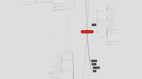
1. Esophagus
1.1. 25cm long
1.2. Narrowest part of alimentary path
1.3. Most muscular segment of GIT
1.4. Typical 4-layered alimentary tube structure
1.5. Non-keratinized stratified squamous epithelium
1.6. Diffuse lymphatics in lamina propria
1.7. Modified subepithelial layers
1.7.1. Muscularis mucosae
1.7.1.1. Longitudinal smooth mm.
1.7.1.2. Thickest in proximal region
1.7.2. Muscularis externa
1.7.2.1. Upper 1/3 is straited muscle
1.7.2.2. Middle third is striated & smooth mm
1.7.2.3. Last third is smooth mm w/ rest of gut
1.8. Has adventitia
1.9. Esophageal Glands
1.9.1. Esophageal glands proper
1.9.1.1. Occur in submucosa
1.9.1.2. Mucous produced by esoph. Glands proper (acidic)
1.9.2. Esophageal cardiac glands
1.9.2.1. Occur in Lamina propria of mucosa
1.9.2.2. Similar to cardiac glands of stomach
1.9.2.3. Present in terminal parts of esophagus
1.9.2.4. Produce neutral mucous
1.10. Barret's esophagus
1.10.1. Metaplasia in cells of the lower end of esophagus
1.10.2. Caused by chronic acid exposure
1.10.3. Normal lining of esophagus (squamous epithelium) replaced by intestinal-type (columnar epithelium)
2. Alimentary canal
2.1. Mucosa
2.1.1. Epithelium
2.1.2. Lamina propria
2.1.2.1. Vascular
2.1.2.1.1. Fenestrated vessels
2.1.2.1.2. Absorption in small & large intestines
2.1.2.1.3. Numerous lymphatic capillaries
2.1.2.2. Contains glands
2.1.2.3. Components of immune system
2.1.3. Muscularis mucosae
2.1.3.1. Forms boundary between mucosa & submucosa
2.1.3.2. Deepest portion of mucosa
2.1.3.3. 2 layers
2.1.3.3.1. Inner circular
2.1.3.3.2. Outer longitudinal
2.2. Submucosa
2.2.1. Moderately dense irregular CT
2.2.2. Larger blood vessels
2.2.3. Lymphatics
2.2.4. Glands in submucosa of esophagus and initial duodenum
2.2.5. Nerve plexus - submucosal plexus (Meissner's plexus) innervate smooth muscle. Post-ganglionic parasympathetic neurons.
2.3. Muscularis Externa
2.3.1. Two layers of smooth mm.
2.3.1.1. Circular layer
2.3.1.2. Longitudinal layer
2.3.2. Contains Myenteric plexus (Auerbach's plexus)- Enteric ANS
2.3.2.1. Hirschsprung Disease (congenital megal colon)
2.3.2.1.1. Genetic mutation
2.3.2.1.2. Arrest in migration of neural crest cells to distal colon
2.3.2.1.3. Absence of enteric nervous system
2.4. Serosa (adventitia)
2.4.1. Membrane - simple squamous epithelium. eg. Mesothelium
2.4.2. Extraperitoneal organs (esophagus, duodenum, ascending colon, descending colon) - absent serosa
3. Stomach
3.1. Layers
3.1.1. Mucosa
3.1.2. Submucosa
3.1.3. Muscularis (3 layers)
3.1.4. Serosa
3.2. Rugae (temporary glands)
3.3. Cardia
3.3.1. Cardiac glands
3.3.1.1. Gastric juice
3.3.1.2. Tubular glands
3.3.1.3. Turtuous
3.3.1.4. Sometimes branches
3.3.1.5. Mucous secreting cells
3.3.1.6. Enteroendocrine cells
3.4. Pylorus
3.4.1. Pyloric glands
3.4.1.1. Branched tubular glands- coiled
3.4.1.2. Neuroendocrine cells
3.4.1.3. Glands empty into deep gastric pits
3.4.1.4. Glands are shorter while gastric pits are longer- opposite of fundic region
3.5. Fundus
3.5.1. Fundic or Gastric glands
3.5.1.1. Produce digestive juice of stomach
3.5.1.2. Simple branched tubular glands
3.5.1.3. Extend from bottom of gastric pits to muscularis mucosae
3.5.1.4. Gastric pits shorter, glands longer
3.5.1.5. Several glands open into one gastric pit
3.5.1.6. Three divisions
3.5.1.6.1. Neck (parietal & mucous cells)
3.5.1.6.2. Base or fundic (chief cells)
3.5.1.6.3. Isthmus (undifferentiated stem cells)
3.6. Simple Columnar mucus cells
3.7. Gastric pits or Foveolae on mucosal surface
3.8. Gastric glands empty into bottom of gastric pits
4. Tongue
4.1. Filiform Papillae
4.1.1. Most numerous & smallest
4.1.2. Conical
4.1.3. Stratified squamous
4.1.4. No taste buds
4.1.5. Anterior dorsal surface of tongue
4.2. Fungiform Papillae
4.2.1. Mushrooom-shaped
4.2.2. Found on dorsal surface
4.2.3. Most numerous near the tip
4.2.4. Taste buds are present
4.3. Circumvallate Papillae
4.3.1. Large, dome-shaped
4.3.2. Anterior to Sulcus Terminalis
4.3.3. 8-12 in humans
4.3.4. Surrounded by moat-like space lined w/ strat. squamous epithelium
4.3.5. Lateral surface has taste buds
4.3.6. Ducts of lingual salivary glands- Von Ebner's Glands
4.3.7. Serous secretions from VE glands clean debris from moat
