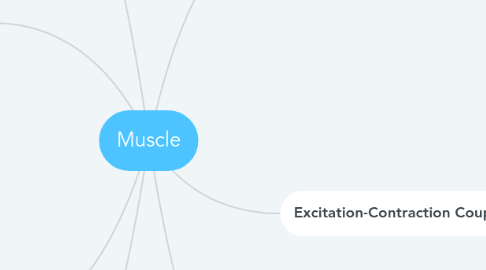
1. Whole Skeletal Muscles
1.1. Cross-bridge activity within muscle fibers accounts for force
1.2. Tension generated by whole muscle is directly related to number of actively contracting muscle fibers.
1.2.1. Amount of tension generated from each muscle fiber is determined by frequency of action potentials from motor neuron
1.3. Speed with which a muscle shortens decreases as the load it lifts increases
1.4. Work performed by a muscle is the product of force produced by the muscle and the distance it shortens.
2. Muscle Energetics
2.1. Contractile activity requires the hydrolysis of ATP to provide energy
2.2. ATP produced through three principal means
2.2.1. 1. transfer of the high energy phosphate from creatine phosphate to ADP
2.2.2. 2. glycolysis
2.2.3. 3. oxidative phosphorylation
2.3. Muscle fibers are classified into different types on basis of their biochemical and metabolic features
2.4. Muscles adapted for extremely rapid contractions typically produce less tension than muscles that contract at slower rates
3. Neural Control of Skeletal Muscle
3.1. Neuromuscular organization of vertebrates is characterized by many non-overlapping motor units
3.1.1. Each controlled by single motor neuron
3.1.2. Each muscle fiber within a motor unit generates action potential that spreads rapidly over entire cell membrane and triggers contractile response.
3.2. Vertebrate tonic fibers usually do not generate action potentials
3.2.1. Each fiber is typically innervated by a single motor neuron that makes multiple synaptic contacts
3.3. The neuromuscular organization of arthropods is characterized by few motor neurons, overlapping motor units, and in some cases, by peripheral inhibition
3.3.1. Each muscle fiber is innervated by more than one motor neuron
4. Vertebrate Cardiac Muscle
4.1. Classified as striated
4.2. Presence of intercalated discs between cells
4.3. Capable of generating endogenous action potentials at periodic intervals.
5. Vertebrate Skeletal Muscle Cells
5.1. Consists of longitudinally arranged muscle fibres
5.1.1. Fibers consist of myofibrils
5.1.1.1. Myofibrils consist of two myofilaments (organized into sacromeres)
5.1.1.1.1. Thick
5.1.1.1.2. Thin
5.1.1.1.3. Filaments slide by each other during contraction
5.1.1.2. Myofibrils consist of sacromeres that make tissue look striated
6. Excitation-Contraction Coupling
6.1. Sarcoplasmic reticulum (SR) sequesters Ca2+ ions to keep cytoplasmic conc. of Ca2+ low
6.1.1. Terminal cisternae of SR possesses RyR calcium channels
6.1.1.1. Transverse tubules include voltage sensitive DHPRs that come into contact with the RyRs of the SR
6.2. Each skeletal muscle contraction is initiated by an action potential.
6.2.1. Gives rise to muscle fiber action potential
6.2.2. Propagates over the cell membrane of the muscle fiber and depolarizes the DHPRs in the t-tubules.
6.2.2.1. Ca2+ ions diffuse out of the terminal cisternae of SR into cytoplasm
7. Vertebrate Smooth (Unstriated) Muscle
7.1. Smooth muscles make up the walls of tubular and hollow organs, and are found in the eye and at the base of hairs and feathers
7.1.1. Contract slowly because their myosin ATPase isomers hydrolyze ATP very slowly
7.1.2. Contraction is controlled by Ca2+
7.1.3. Relaxation occurs when cytoplasmic Ca2+ decreases

