

1. Basics
1.1. Capsular pattern
1.1.1. Limited F/ I.R., Some limitation in Abd, No or lil Limited Add and ER
1.2. close pack position
1.2.1. Ligamentous: Full ext, abd, IR
1.2.2. Bony: 90 deg F, Slight Abd., Slight ER
1.3. loose pack position
1.3.1. 30 deg F, 30 deg Abd., Slight LR
1.4. Roll and slide: Opposite
1.5. ROM
1.5.1. Flexion: 120
1.5.2. Extension: 15
1.5.3. Abduction: 45
1.5.4. Adduction: 30
1.5.5. Internal Rotation: 45
1.5.6. External Rotation: 45
2. Joint Conditions
2.1. Osteoarthritis
2.1.1. Classification Criteria
2.1.1.1. 1. Hip pain
2.1.1.2. (2a) Hip IR <15 (2b) ESR <= 45mm/hr or HF <115 deg
2.1.1.3. 3a Hip IR >=15 3b Pain on Hip IR
2.1.1.4. 3c Morning stiffness of hip >60 min 3d age>50 yrs
2.1.2. HD heberden nodes
2.1.3. Combined criteria : Hip pain + any 2 of fall, ESR <=20mm/hr, osteophytes, reduced joint space
2.1.4. Test: Hip compression test, Scouring
2.1.5. Rx: Initially open chain because weight bearing pain, aquatic exercise
2.2. Rheumatoid Arthritis
2.2.1. morning stiffness
2.2.2. Criteria: Synovitis in atleast 1 joint, duration of symptoms
2.3. Avascular necrosis
2.3.1. FAbIR problem (Loss)
2.3.2. Coxalgic gait
2.3.3. Symptoms: Groin pain, thigh, tenderness on palpation
2.3.4. Tx Joint protection, no cross sitting
2.4. Splint: Trilateral, Pavlic harness, Frejka pillow, Von Rosen splint
2.5. anteverted hip
2.5.1. At birth around 30 deg, adult around 8-15 degree
2.5.2. Squinting patella, apex MR
2.5.3. Toe in
2.5.4. Test:
2.5.4.1. Craig Test: b/w femoral neck & condyle
2.5.4.2. Pigeon toes
2.5.4.3. Excess MR, reduced LR in extension
2.6. coxa vara
2.6.1. neck shaft angle <120
2.7. coxa valga
2.7.1. neck shaft angle >135
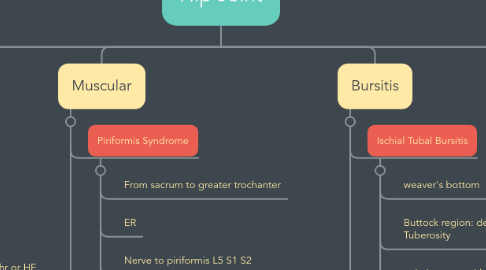
3. Muscular
3.1. Piriformis Syndrome
3.1.1. From sacrum to greater trochanter
3.1.2. ER
3.1.3. Nerve to piriformis L5 S1 S2
3.1.4. Test: Piriformis Test
3.2. IT band friction disorder
3.2.1. Lateral knee pain
3.2.2. In athletes
3.2.3. Test: Noble compression test (pain at 30 deg F)
3.2.4. Tx Taping, reduce activity, MFR
3.3. IT band tightness
3.3.1. Ober test
3.3.2. Modified ober test: Conditions like recent TKR
3.3.3. Prone lying test
3.3.4. Tx Stretching
4. Bursitis
4.1. Ischial Tubal Bursitis
4.1.1. weaver's bottom
4.1.2. Buttock region: deep b/w Gmax & Ischial Tuberosity
4.1.3. pain increase with prolong sitting, may radiate to leg
4.1.4. swelling & limited mobility
4.1.5. Test: Sign of the buttock (SLR +kf, Hip ROM
4.1.6. Tail bone pain
4.2. Trochanteric Bursitis
4.2.1. Subgluteal medius bursa and subgluteus maximus bursa
4.2.2. snap felt at lateral aspect of hip
4.2.3. pain on palpation
4.2.4. Activity related pain: Pain in PROM, AROM
4.2.5. Management: Reduce or modify activity, cryotherapy, TENS, Taping
4.2.6. anterolateral pain in AROM, PROM
4.2.7. pain ascending stairs
4.2.8. chronic pain, tenderness
5. Pediatric condition
5.1. Hip dysplasia
5.1.1. Test:
5.1.1.1. Bryant's triangle
5.1.1.1.1. Abduction Test (Hart sign)
5.1.1.1.2. Galeazzi Sign (1 knee high)
5.1.2. Barlow
5.1.2.1. Close book
5.1.2.1.1. Hip flexion 90 deg, Knee flexion, Abduction
5.1.3. Ortolani
5.1.3.1. open book
5.1.3.2. Hip flexion, Abd, pull anteriorly
5.1.3.3. Fix the dislocation
5.2. LCPD
5.2.1. H/o osteonecrosis
5.2.2. 2-13 YRS
5.2.3. Psoatic limp
5.2.4. Limb presentation F AD ER
5.2.5. Cresent sign in X ray
5.2.6. No blood supply
5.2.7. Tx: Joint protection, splint (patten bottom, toronto) FABER stability position
5.3. SCFE
5.3.1. 10-17 yrs
5.3.2. obese, sedantry, vague pain
5.3.3. limit FABIR
5.3.4. Trendelenberg gait
5.3.5. Imaging: Gluteal fold disappear
5.3.6. Displaced femoral epiphysis
5.3.7. Tx: Education, stability, splint, Intramedullary nailing
6. other
6.1. meralgia paresthetica
6.1.1. lateral femoral cutaneous nerve involved
6.1.2. entrapment at level of inguinal ligament
6.1.3. L2 L3
6.1.4. Pain, burning, numbness at anterolateral thigh
6.1.5. pain decreases with sitting due to decrease in tension on ligaments
6.1.6. Test: NCV
6.1.7. Tx:
6.1.7.1. Wear loose clothing
6.1.7.2. Losing excess weight
6.2. impingement
6.2.1. Anteroposterior Impingement Test
6.2.1.1. For retroversion, SCFE, Femoroacetabulat impingement
6.2.1.2. Hip Flexion 90 then do MR + Adduction
6.2.2. Posteroinferior Impingement Test
6.2.2.1. Hip extension then Lateral Rotation
6.2.3. Femoroacetabular Impingement
6.2.3.1. Mc Carthy Test
6.2.3.2. C sign
6.3. Femoral Neck stress fracture
6.3.1. Test:
6.3.1.1. Fulcrum Test of hip
6.3.1.2. Heel strike Test
6.3.1.3. Patellar Pubic Percussion Sign: dull sound