
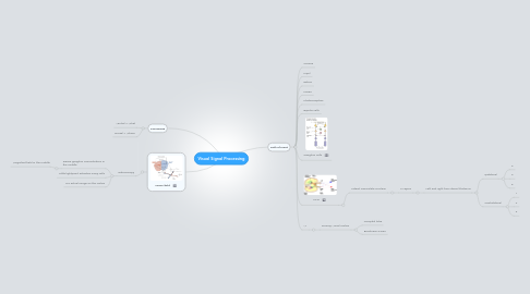
Visual Signal Processing
af Mentor Palokaj
1. Processing
1.1. Ventral = What
1.2. Dorsal = Where
2. Vision field
2.1. Retinoscopy
2.1.1. Dense ganglion concentration in the middle
2.1.1.1. Magnified field in the middle
2.1.2. Little lightpoint activates many cells
2.1.3. No actual image on the cortex
3. Path of input
3.1. Cornea
3.2. Pupil
3.3. Retina
3.4. Fovea
3.5. Photoreceptors
3.6. Bipolar cells
3.7. Ganglion cells
3.8. LGN
3.8.1. Lateral Geniculate Nucleus
3.8.1.1. 6 Layers
3.8.1.1.1. Left and right from dorsal thalamus
3.9. V1
3.9.1. Primary Visual Cortex
3.9.1.1. Occipital lobe
3.9.1.2. Brodmann's area