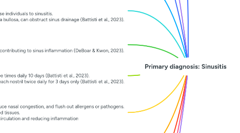
1. Causative Factors Viral factors: Rhinovirus and influenzae (DeBoer & Kwon, 2023). Bacterial factors: Streptococcus pneumoniae, H. Influenzae, and M. Catarrhalis (DeBoar & Kwon, 2023). Environmental factors: air pollution and tobacco smoke (DeBoar & Kwon, 2023). Anatomical factors: deviated nasal septum, nasal polyps, and concha bullosa (DeBoar & Kwon, 2023).
2. Clinical manifestations: The symptoms of sinusitis include nasal congestion and discharge, which is often purulent. Patients typically experience facial pain or pressure, particularly in the forehead, cheeks, or around the eyes, which tends to worsen when bending forward. A reduced sense of smell (hyposmia or anosmia) is also frequent. Cough, often more pronounced at night due to postnasal drip, is another common complaint. Fever may accompany these symptoms, especially in acute bacterial sinusitis. Patients often report fatigue and a general sense of malaise (Battisti et al., 2023).
3. Objective Findings: Swollen and erythematous nasal mucosa, often with visible purulent discharge. Tenderness on palpation over the sinuses, particularly the frontal and maxillary areas, can indicate sinus involvement. Examination of the oropharynx could show postnasal drip (Battisti et al., 2023).
4. Risk Factors: Upper respiratory infections, like the common cold. Allergies, particularly allergic rhinitis, can cause mucosal swelling and predispose individuals to sinusitis. Anatomical variations, such as a deviated nasal septum, nasal polyps, or concha bullosa, can obstruct sinus drainage (Battisti et al., 2023).
5. Demographics: Sinusitis affects 16% of women and 10% of men each year (Jnjmedtech, 2019).Diagnostic: CT Scan of the Sinuses: This is the gold standard imaging technique for diagnosing sinusitis (Battisti et al., 2023). Allergy test: in cases of chronic or recurrent sinusitis, allergy testing may be conducted to identify any underlying allergic triggers that could be contributing to sinus inflammation (DeBoar & Kwon, 2023).
6. Pharmacological intervention Amoxicillin 500 mg orally three times daily 10 days (Battisti et al., 2023). Oxymetazoline 2 sprays into each nostril twice daily for 3 days only (Battisti et al., 2023).
7. Non-pharmacological Saline irrigation: Regularly flushing the nasal passages with saline solution can help clear mucus, reduce nasal congestion, and flush out allergens or pathogens. Inhaling steam from hot water to moisten the nasal passages, reduce congestion, and soothe irritated tissues. Applying a warm compress to the face can help relieve sinus pain and pressure by improving blood circulation and reducing inflammation Sleeping with the head elevated can help facilitate sinus drainage and reduce congestion.
8. Patient education (DeBoar & Kwon, 2023) Rest No smoking Sleep with head elevated Avoid allergies if possible More water intake
9. Referrals May refer to Otolaryngologist if symptoms do not get better after 3 months of treatment (DeBoar & Kwon, 2023).
10. Red Flags Cranial nerve abnormalities, especially changes in eye shape, vision changes, or eye pain Worsening headache Fever that won't go away
11. Patho: Characterized by the inflammation of the mucosal lining of the paranasal sinuses. This condition often begins with an obstruction of the sinus ostia, the small openings that allow mucus to drain from the sinuses into the nasal cavity (Battisti, et al., 2023).