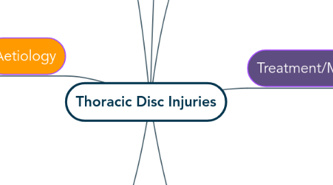
1. Epidemiology
1.1. Indications of yellow flags for Thoracic Disc injuries are in patients that may be smokers, overweight or obese, engage in a sedentary lifestyle, and have a poor level of fitness
1.2. Most commonly occur in patients in their mid to late adult life due to degeneration (Degenerative disease patients)
1.3. Trauma
1.4. Sports involving axial rotation of the spine can increase the risk of thoracic disc injuries. Eg. Golf
2. Aetiology
2.1. Thoracic disc injury is most commonly due to degeneration of the disc
2.2. There are a number of different types of thoracic disc injuries
2.2.1. Disc Herniation
2.2.2. Annular Tear: Tearing of the Annulus Fibrosis due to insufficient distribution of compressive loads across the disc
2.2.3. Disc Degeneration: With age, the disc becomes less hydrated resulting in less elasticity and energy storage. It also results in an impaired ability of the disc to repair itself,
2.2.4. Degenerative changes in the thoracic spine can damage the intervertebral disc. Eg. Bone spurs
2.3. Thoracic disc injuries can also be caused by repeated micro traumas over time, usually of a twisting or torsional nature, as seen in Golf. It may also be due to an excessive mechanical loading or an acute trauma to the intervertebral disc (IVD).
3. Clinical Features
3.1. Pure discogenic pain may be dull and localised to the region.
3.2. The location and quality of pain is largely dependent on the type of disc injury and if there is any compromisation of neural tissues
3.3. It is also possible for upper/lower thoracic disc herniation’s to manifest as cervical or lumbar pain
3.4. Correct diagnosis is important as pain may be referred to the retrogastric, retrosternal, or inguinal areas, resulting in misdiagnoses such as Cholecystitis, Myocardial Infarction, Hernia, or Nephrolithiasis
4. Prognosis/Natural Healing
4.1. Thoracic Disc injuries are usually self limiting. Most cases can resolve with treatment within 4-6 weeks following onset.
4.2. Surgical interventions are becoming increasingly more effective and safe for patients with thoracic disc injuries
4.2.1. The most serious but rare complication of thoracic disc disorder is myelopathy. Surgical intervention is indicated in ca
5. Treatment/Management
5.1. Gentle Osteopathic treatment such as articulation and soft tissue of the surrounding structures. Any correction of imbalances in the body may also be performed to reduce any additional load on the injured area
5.2. Rehabilitation is important, especially in athletes. Strengthening of the dynamic stabilisers of the spine is important to counteract any significant forces on the spine.
5.3. In severe cases, Epidural cortisone injections or Surgical Intervention may be indicated
5.3.1. The most serious but rare complication of Thoracic Disc Injuries is Myelopathy. Surgical decompression is indicated in cases of worsening Myelopathy, however there is a chance the patient will not improve
5.4. Education of the patient regarding proper spinal mechanics and spinal stabilisation.
6. Examination Procedure
6.1. Observation of the patient, especially of the thoracic region. (Look for any TART findings) Active, Passive and Active Resisted Range of Motion testing, accompanied by palpation for any anomalies/abnormalities
6.2. Clinical testing such as springing of the ribs, maximal inhalation, cough/sneeze test Slump test -> neural tissue provocation
6.3. Referral for imaging is sometimes indicated.
6.3.1. MRI may be used to detect any disc abnormalities, however it cannot determine the size and location or characterise a herniation.
6.3.1.1. MRI may be used to detect any disc abnormalities, however it cannot determine the size and location or characterise a herniation.
6.3.1.1.1. New node
6.3.2. Usually an X-Ray of the Thoracic Spinal Region contributes little to the clinical picture unless detection of conditions such as Scheurmanns or the presence of a Secondary neoplastic disorder is required.

