

1. Specimen
1.1. Depends on the type of infection. (Pus, joint aspirate, blood, sputum, stool. Urine ).
2. Culture
2.1. Based on their consistency a) solid medium:– contains 2% agar Colony morphology, pigmentation, hemolysis can be appreciated. Eg: Nutrient agar, Blood agar b) liquid medium:– no agar. For inoculum preparation, Blood culture, for the isolation of pathogens from a mixture. Eg: Nutrient broth c) semi solid medium:– 0.5% agar. Eg: Motility medium
2.2. Based on the constituents/ ingredients a) simple medium b) complex medium c) synthetic or defined medium d) Special media:-
2.2.1. Enriched media: Substances like blood, serum, egg are added to the basal medium. Used to grow bacteria that are exacting in their nutritional needs. Eg: Blood agar, Chocolate agar
2.2.2. Enrichment media: Liquid media used to isolate pathogens from a mixed culture. Media is incorporated with inhibitory substances to suppress the unwanted organism.
2.2.3. Selective media: The inhibitory substance is added to a solid media. Eg: Mac Conkey’s medium for gram negative bacteria
2.2.4. Indicator media: These media contain an indicator which changes its colour when a bacterium grows in them.
2.2.5. Differential media: A media which has substances incorporated in it enabling it to distinguish between bacteria. Eg: Mac Conkey’s medium Distinguish between lactose fermenters(pink) & non lactose fermenters.
2.2.6. Sugar media
2.2.7. Transport media
2.2.8. Media for biochemical reactions
2.3. Based on Oxygen requirement - Aerobic media - Anaerobic media: use Anaerobic
2.4. Capnophiles require high CO2: use -Candle jar - CO2-packet
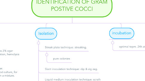
3. Isolation
3.1. Streak plate technique: streaking.
3.1.1. pure colonies
3.2. Slant inoculation technique: dip & zig zag.
3.3. Liquid medium inoculation technique: scrath well
3.4. Semisolid medium inoculation technique: dip
3.4.1. Motile bacteria and NON-motile bacteria
4. incubation
4.1. optimal tepm. 24h at 37ْ
5. Inspection
5.1. Macroscopy : size, shape..
5.2. Microscopy : Smear prepration
5.2.1. loop full of distilled water (in solid media) pick a colony and mix drying Fix
5.2.1.1. Gram staining: Fix by methanol Crystal violet (primary stain) Gram’s iodine (mordant) Acetone-alcohol (decolorizing agent) Safranin (counter stain)