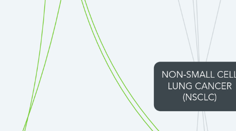
1. Treatments
1.1. Varies by stage of cancer
1.2. Surgery to remove cancerous tissue
1.2.1. Option for early stages
1.2.1.1. Pneumonectomy: removes entire lung
1.2.1.2. Lobectomy: entire lobe is removed
1.2.1.2.1. Preferred type if possible
1.2.1.3. Segmentectomy/Wedge resection: only part of lobe removed
1.2.1.4. Sleeve Resection: cancerous section is cut out, two ends are sewn back together
1.3. Radiofrequency Ablation (RFA)
1.3.1. High-energy radio waves heat the tumor and destroys cancerous cells
1.4. Radiation Therapy
1.4.1. Targeted radiation used to disrupt cancer growth
1.4.1.1. Main treatment (when surgery is not an option)
1.4.1.2. Shrinking the tumor before surgery/removing areas after surgery
1.4.1.3. Treat cancer spread
1.4.1.4. Palliative
1.5. Chemotherapy
1.5.1. Attacks fast-growing cells
1.5.2. Often combination of two anti-cancer drugs
1.5.2.1. Cisplatin or carboplatin
1.5.2.2. Gemcitabine with vinorelbine or paclitaxel
1.5.3. Given in cycles
1.6. Targeted Drug Therapy
1.6.1. Angiogenesis
1.6.2. EGFR changes
1.6.3. T790M mutation
1.6.4. ALK gene changes
1.6.5. BRAF gene changes
1.7. Immunotherapy
1.7.1. Boost the immune response to cancerous tissues
1.8. Palliative Treatments
1.8.1. Treat fluid build-up
1.8.1.1. Heart
1.8.1.2. Lungs
1.8.2. Airway blocked by a tumor
2. Signs/Symptoms
2.1. Earlier Stages
2.1.1. Persistent or worsening cough
2.1.2. Coughing up blood/rust-colored sputum
2.1.3. Chest pain
2.1.3.1. Worsens with
2.1.3.1.1. Deep breathing
2.1.3.1.2. Coughing
2.1.3.1.3. Laughing
2.1.4. Hoarseness
2.1.5. Weight loss/loss of appetite
2.1.6. Shortness of breath
2.1.7. Feeling tired or weak
2.1.8. Persistent infections
2.1.8.1. Bronchitis
2.1.8.2. Pneumonia
2.1.9. New onset of wheezing
2.2. Paraneoplastic Syndromes
2.2.1. Interfere with diagnosis
2.2.1.1. Hypercalcemia
2.2.1.1.1. Frequent urination/thirst
2.2.1.1.2. Constipation, nausea, vomiting, belly pain
2.2.1.1.3. Weakness, fatigue, dizziness, confusion, other NS problems
2.2.1.2. Excess growth or thickening of bones
2.2.1.2.1. Fingertips especially
2.2.1.2.2. Often painful
2.2.1.3. Blood clots
2.2.1.3.1. New node
2.2.1.4. Gynecomastia
2.2.1.4.1. Excess breast tissue in men
2.3. Metastatic S/Sx
2.3.1. Bone pain
2.3.2. Nervous system changes
2.3.2.1. Headache
2.3.2.2. Weakness/numbness of limbs
2.3.2.3. Dizziness
2.3.2.4. Balance problems
2.3.2.5. Seizures
2.3.3. Jaundice (yellowing of skin or eyes)
2.3.3.1. Spread to liver
2.3.4. Lumps near surface of body
2.3.4.1. Cancer spread to skin or lymph nodes
3. Incidence/Prevalence
3.1. Smokers have a much higher incidence rate
3.2. Overall Numbers (include smokers and non-smokers)
3.2.1. 1 in 14 men
3.2.1.1. African-American males 20% more likely than white males
3.2.2. 1 in 17 women
3.2.2.1. African-American women 10% less likely than white women
3.3. Leading cause of cancer death among both men and women
3.3.1. “1 out of 4 cancer deaths are from lung cancer”
3.4. Occurs mostly in older individuals
3.4.1. 66% of those diagnosed >65 y/o
3.5. Around 230,000 new cases in 2017
3.6. 80-85% of lung cancers are NSCLC
4. Pathogenesis
4.1. Uncontrolled cell proliferation in different areas of tissues (specifically lungs here) due to any number of damaging factors and alack of proper tumor suppressor genes
4.1.1. Lung cancer tends to occur due to some kind of chronic inflammation causing cellular DNA damage, leading to uncontrolled proliferation, can be caused by genetic factors which may lead to a lack of tumor suppressor and/or caretaker genes
4.1.1.1. Tumor eventually becomes vascularized through angiogenesis and as it grows, can be categorized in stages. The tumor can become metastatic, meaning it will release cells into the blood stream that travel to nearby and distant sites and create new growths
4.1.1.1.1. Stage 0: cancer is found only in top layers of cells lining the bronchi, has not invaded deeper tissue or metastasized
4.1.1.1.2. Stage IA: tumor <3 cm across, has not reached the visceral pleura or the main bronchi. No metastasis to lymph or distant sites
4.1.1.1.3. Stage IB: has one or more:
4.1.1.1.4. Stage IIA: 3 categories
4.1.1.1.5. Stage IIB
4.1.1.1.6. Stage IIIA
4.1.1.1.7. Stage IIIB
4.1.1.1.8. Stage IV
5. Diagnostics
5.1. Imaging Tests
5.1.1. Chest X-ray
5.1.1.1. Often the first performed
5.1.2. Computed tomography (CT) scan
5.1.2.1. Shows size, shape, position of tumors
5.1.2.2. CT-guided needle biopsy
5.1.2.2.1. CT scan can be used to guide biopsy needle into suspected area
5.1.3. Magnetic resonance imaging (MRI) scan
5.1.3.1. Often used to look for metastasis
5.1.4. Positive emission tomography (PET) scan
5.1.4.1. Can be used with CT scan
5.1.4.2. Used to look for spread to lymph nodes and evaluate treatment
5.1.5. Bone scan
5.1.5.1. Radioactive material injected into blood
5.1.5.1.1. Can show metastasis to bones
5.2. Lung-cancer Specific Testing
5.2.1. Sputum Cytology
5.2.1.1. Sputum is examined under microscope for cancer cells
5.2.1.2. More likely to find squamous cell or others that start in major airways in the lung
5.2.1.2.1. Other testing is required
5.2.2. Thoracentesis
5.2.2.1. Pleural effusion (build-up of fluid around the lungs) is removed
5.2.2.1.1. Fluid is checked for malignant cells
5.2.2.2. Can be repeated to remove fluid if malignant pleural effusion is found
5.2.3. Needle Biopsy
5.2.3.1. Fine Needle Aspiration (FNA)
5.2.3.1.1. Very thin needle removes cells and fragments of tissue for microscopic examination
5.2.3.2. Core biopsy
5.2.3.2.1. Larger needle removes core sample of tissue
5.2.3.3. Transthoracic needle biopsy
5.2.3.3.1. Outer part of lungs suspected
5.2.4. Bronchoscopy
5.2.4.1. Can find tumors or blockages in larger airways
5.2.4.1.1. Biopsies during procedure
5.2.4.1.2. Bronchoscope passed down trachea with a lighted tube and camera
5.3. Detecting Spread - Chest
5.3.1. Endobronchial ultrasound
5.3.1.1. Transducer passed down trachea to inspect lymph nodes and other areas between the lungs
5.3.1.2. Samples can be taken
5.3.2. Endoscopic esophageal ultrasound
5.3.2.1. Very similar procedures
5.3.2.2. Samples can be taken
5.3.3. Mediastinoscopy
5.3.3.1. Small cut made in front of neck and thin, hollow lighted tube inserted behind the sternum
5.3.3.1.1. Samples can be taken and examined from lymph nodes, trachea, and major bronchial tube areas
5.3.4. Mediastinotomy
5.3.4.1. Slightly larger incision between left second and third ribs next to the breast bone
5.3.4.1.1. Reach harder-to-reach lymph nodes for biopsy
5.3.5. Thorascopy
5.3.5.1. Done specifically for chest wall spread
5.3.5.1.1. Operating room, under anesthesia
5.4. Lab Testing on Samples
5.4.1. Immunohistochemical Tests
5.4.1.1. Thin slices of sample are subjected to antibodies which bind to cancer cells; chemicals are added which change antibody colors
5.4.2. Molecular tests
5.4.2.1. Specific genetic changes might help drive treatment
5.4.2.1.1. The Epidermal Growth Factor (EGFR)
5.4.2.1.2. ALK gene changes in light or non-smokers
5.4.2.1.3. BRAF gene changes
5.4.3. Blood tests
5.4.3.1. Complete Blood Count
5.4.3.1.1. Directs treatment
6. Etiology/Risk Factors
6.1. Tobacco smoke
6.1.1. second-hand smoke exposure can also be a factor
6.2. Radon exposure
6.3. Workplace Exposures
6.3.1. Asbestos
6.3.2. Radioactive agents such as uranium
6.3.3. Diesel exhaust
6.3.4. Arsenic
6.3.4.1. Also found in drinking water in less-developed countries
6.4. Previous radiation therapy
6.4.1. Especially targeted to the chest
6.5. Air pollution
6.6. Family history of lung cancer
6.6.1. Could be related to
6.6.1.1. Genetics
6.6.1.1.1. Inherited gene changes
6.6.1.1.2. Acquired gene changes
6.6.1.2. Family environmental factors
