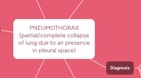
1. Pathogenesis
1.1. Classifications
1.1.1. SPONTANEOUS- occurs w/out obvious injury cause; rupture of air-filled blub/bullae on lung surface results in atmospheric air into pleural cavity; aveolar pressure>pleural pressure so negative pressure causes lung to collapse from recoil. 2 types:
1.1.1.1. primary- occurs in the healthy
1.1.1.2. secondary- individuals w/ underlying lung disease
1.1.2. TRAUMATIC
1.1.2.1. penetrating or blunt chest injuries to pleura
1.1.2.1.1. fractured trachea/bronchus/ribs OR ruptured esophagus OR medical procedures (i.e. central line insertion, chest compressions)
1.1.3. TENSION
1.1.3.1. chest/respiratory injury permits air entry but does not allow to exit; intrapleural pressure>Atmospheric pressure
1.1.3.1.1. compression of vena cava & atelactasis (unaffected lung), mediastinum shift all reduce stroke volume
1.1.3.1.2. intrapleural pressure lowers cardiac contraction = reduced stroke volume
1.1.3.1.3. mechanical ventilation use due to underlying lung disease can be a risk factor
2. Diagnosis
2.1. Chest radiograph
2.2. CT scans
2.3. pulse oximetry
2.4. blood gas analysis
3. Treatment
3.1. SMALL SPONTANEOUS
3.1.1. Prevention: lifestyle (smoking cessation, avoiding high altitude exposure, scuba diving, non-pressurized aircraft)
3.1.2. Interventions: follow-up observation, chest radiographs, supplemental 02
3.2. LARGE PNEUMOTHORAX
3.2.1. air removal via closed-chest tube drainage system (w/ OR w/out suction)
3.3. TRAUMA/EMERGENCY
3.3.1. continuous chest suction- large bore needle into affected area allows in & out a/e to promote lung re-expansion; heimlich valve/chest tube must be inserted immediately after
4. Nursing Interventions
4.1. Assess chest expansion, observe pallor
4.2. Monitor vitals (HR, RR, SpO2)
4.3. Assess bilateral air entry via chest auscultation
4.4. Assess subcutaneous emphysema
4.5. Adjust HOB to Fowler's
4.6. Administer O2 therapy as ordered
4.7. Provide wound care for open pneumothorax injury
4.8. Maintain chest-tube system for pts.
5. Risk Factors/Prognosis
5.1. Age: 20-40 years
5.2. Men>Women
5.3. Build: Tall and underweight
5.4. Lifestyle: Smoking, Lung Disease Hx, mechanical ventilation use
5.5. Genetics
5.6. Hx of prior pneumothorax ^ risk of recurrence within 1-2 years
5.7. severe hypoxemia & hypotension in extreme cases; most common to recover from 1-2 weeks
6. Clinical Manifestations
6.1. SPONTANEOUS
6.1.1. ipsilateral chest pain, dyspnea, chest asymmetry, hyperresonance sounds, decreased/absent breath sounds in affected area
6.1.1.1. Primary VS. Secondary
6.1.1.1.1. blood flow shift to unaffected lung due to hypoxemic vasoconstriction easily resolved in primary pts. but is critical in secondary pts. due to their lack of compensation ability for the ^HR & SV
6.2. TRAUMATIC
6.2.1. hypoxemia, O2 desaturation, hypotension (in severe cases)
6.3. TENSION
6.3.1. mediastinal shift, ^ intrathoracic pressure = decreased SV = decreased CO and ^HR, neck vein distension, subcutaneous emphysema, shock
