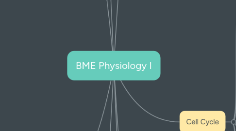
1. Cell Signaling
1.1. Pathways
1.1.1. Juxtacrine (contact dependent)
1.1.2. Paracrine/autocrine
1.1.3. Synaptic
1.1.4. Endocrine
1.2. Classes
1.2.1. G-Protein-Coupled Receptors
1.2.1.1. Structure
1.2.1.1.1. Trimeric
1.2.1.1.2. 7 transmembrane segments
1.2.1.2. Function
1.2.1.2.1. GAP
1.2.1.2.2. GEF
1.2.1.3. Examples
1.2.1.3.1. Cyclic AMP production
1.2.1.3.2. GPCR>IP3>Calcium
1.2.2. Ion-Channel Coupled Receptors
1.2.3. Enzyme-Coupled Receptors
1.2.3.1. Structure
1.2.3.1.1. ligand bind on outer surface
1.2.3.1.2. 1 transmembrane segment
1.2.3.1.3. scaffold on cell membrane
1.2.3.2. Examples
1.2.3.2.1. Receptor Tyrosine Kinase
1.2.3.2.2. MAP Kinase
1.2.3.2.3. Rho
1.2.3.2.4. JAK-STAT
1.2.3.2.5. TGF Beta/SMAD
1.2.3.2.6. PI3-Kinase
1.2.4. Regulated Proteolysis
1.2.4.1. NFkB
1.2.4.1.1. inflammation
1.2.4.2. Wnt
1.2.4.2.1. cell growth and proliferation
1.2.4.3. Notch
1.2.4.3.1. pattern formation of tissues during development
1.3. Responses
1.3.1. Sigmoidal
1.3.2. All-or-none
1.3.3. Positive Feedback
1.3.4. Negative Feedback
1.3.4.1. Adaptation/desensitization
2. Cell Membrane
2.1. Lipid Bilayer
2.1.1. Define boundaries
2.1.2. cellular bounds
2.1.3. Organize complex reactions
2.1.4. Regulate information
2.2. Membrane Proteins
2.2.1. Transport
2.2.1.1. Passive - with gradient
2.2.1.2. Active - against gradient
2.2.2. integral
2.3. Cholesterol
2.3.1. Transport
2.3.1.1. HDL + apoA1
2.3.1.2. LDL + apoB
3. Cell Metabolism
3.1. Reactions
3.1.1. Only proceed with -dG
3.1.2. Enzymes lower activation energy
3.1.3. Reactions can be coupled
3.1.4. Reduction - loss of electrons
3.1.5. Oxidation - gain of electrons
3.1.6. Head polymerization - carries bond for addition of next monomer - proteins/fatty acids
3.1.7. Tail Polymerization - carries bond for own addition - DNA, RNA, polysaccharides
3.2. Energy Carriers
3.2.1. ATP
3.2.2. NAD/NADH - oxidation agent (easily reduced)
3.2.3. NADPH - reduction agent (easily oxidized)
3.3. Glycolysis
3.3.1. Glucose split to 2 pyruvate
3.3.2. 2 ATP used
3.3.3. 4 ATP, 2 NADH gained
3.3.4. doesn't use oxygen
3.4. Krebs Cycle
3.4.1. Takes place in Mitochondria
3.4.2. Starts with Acetyl CoA made from pyruvate or fatty acids
3.4.3. Produces 3 NADH, 1 FADH, 1 GTP
3.4.4. Uses oxygen
3.5. Electron transport chain
3.5.1. 30 ATP created
3.5.2. Proteins embedded in inner mitochondrial membrane - many due to cristae
3.5.3. Protons pumped into intermembrane space
3.5.4. ATP Synthase uses electrochemical gradient to make ATP
3.5.4.1. reversible
3.5.4.2. rotor and stator shape
3.5.5. Protons from NADH supplied
4. Cell Junctions
4.1. Cell-Cell
4.1.1. Cadherins
4.1.1.1. low-affinity, high volume binding
4.1.1.2. Signal transduction
4.1.1.3. adhesion belt spanning many cells
4.1.1.4. Catenin links to actin
4.1.1.5. Utilizes actin filaments
4.1.2. Desmosomes
4.1.2.1. mechanical strength
4.1.2.2. utilizes intermediate filaments
4.1.3. Tight Junctions
4.1.3.1. seal between cells
4.1.3.2. permeable to ions and small molecules
4.1.3.3. made with claudins
4.1.3.4. separate apical from basal sides
4.1.3.5. especially present in epithelial cells
4.1.4. Gap-Junctions
4.1.4.1. bridge gap between cells
4.1.4.2. 6 connexins on either side
4.1.4.3. allow ions and small water-soluble molecules to pass
4.2. Other
4.2.1. Selectins
4.2.1.1. All WBCs to roll
4.2.2. Immunoglobins
4.3. Cell-ECM
4.3.1. Integrins
4.3.2. Hemidesmosomes
4.3.2.1. intermediate filaments to ECM
5. Cytoskeleton
5.1. Actin Fliaments
5.1.1. locomotion
5.1.1.1. Together with Myosin II
5.1.1.1.1. contraction
5.1.1.1.2. rapid response
5.1.1.1.3. activated by calcium
5.1.2. cell shape
5.1.3. treadmilling
5.1.4. 6 nm
5.1.5. helical
5.1.6. Binding proteins
5.1.6.1. Monomer availability
5.1.6.2. polymerization acceleration
5.1.6.3. filament dynamics
5.1.6.4. Depolymerization
5.1.6.5. mechanical properties and signaling
5.2. Intermediate Filaments
5.2.1. mechanical strength
5.2.2. ropelike, 10 nm
5.2.3. helical grouping
5.3. Microtubules
5.3.1. hollow
5.3.2. mitotic spindle
5.3.3. 25 nm
5.3.4. Position of organelles
5.3.5. nucleated from MT organizing centers, centrosome
6. Cell Cycle
6.1. Phases
6.1.1. Interphase
6.1.1.1. S
6.1.1.2. G1/G2
6.1.1.3. lasts much longer than M
6.1.1.4. G0 - static phase
6.1.2. M
6.1.3. Decision points
6.1.3.1. G1-S decision to "start"
6.1.3.2. G2-M decision to start M
6.1.3.3. Metaphase-Anaphase decision to split, cytokinesis
6.2. Control
6.2.1. Cyclin-Dependent Kinases
6.2.1.1. cyclins
6.3. Death
6.3.1. Apoptosis
6.3.2. necrosis
6.3.3. Regulated by caspases
6.3.4. Pathways
6.3.4.1. Extrinsic
6.3.4.1.1. Cell-surface death receptors
6.3.4.1.2. Fas activation
6.3.4.2. Intrinsic
6.3.4.2.1. depends on mitochondria
6.3.4.2.2. Cytochrome C, component of electron transport chain
6.3.4.2.3. Bcl2
6.4. transcription
6.4.1. regulators - promotors and repressors
6.4.2. Post transcription control
6.4.2.1. transcription attenuation
6.4.2.2. riboswitches
6.4.2.3. RNA editing
6.4.2.4. Regulated Nuclear transport
6.4.2.5. cytosol localization
6.4.2.6. RNA stability
6.5. Differentiation
6.5.1. DNA methylation
6.5.1.1. can last between cell splits
6.5.1.2. cell "memory"
7. Immune System
7.1. Innate
7.1.1. Granulocytes
7.1.1.1. basophils
7.1.1.2. eosinophils
7.1.1.3. neutrophils
7.1.2. Mast Cells
7.1.3. Monocytes
7.1.3.1. macrophages
7.1.3.2. dendritic
7.1.4. Natural Killer
7.1.5. Compliment Proteins
7.2. Adaptive
7.2.1. B Cells
7.2.2. Antibodies
7.2.3. T Cells
7.2.3.1. CD8+
7.2.3.2. CD4+
7.2.3.2.1. T-helper
7.2.3.2.2. T-reg
8. ECM
8.1. Fibroblasts build ECM
8.2. Glycosaminoglycans (GAGs)
8.2.1. Hyaluronon
8.2.2. Proteoglycans
8.3. Fibrous Proteins
8.3.1. Collagen
8.3.1.1. Type 1, 90% of body's collagen
8.4. Non-collagen Glycoproteins
8.4.1. elastin (arteries)
8.5. Fibronectin
8.6. Basal Lamina
8.6.1. 40-120 nm
8.6.2. flexible sheet
8.6.3. Laminin and Type IV Collagen

