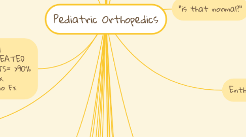
1. pediatric bones
1.1. Growth plates:
1.1.1. ossification center
1.1.2. Germinal matrix
1.1.3. zone of hypertrophy
1.1.4. zone of calcification
1.1.5. zone of ossification
1.2. higher remodeling potential
1.3. increased vasculature
1.4. periosteum strongest element
1.5. growth plate weakest element
1.6. higher risk of diskitis & hematogenous osteomyelitis
2. Fx principles
2.1. peds 10-12 degrees/year remodeling (1 degree/month)
2.2. remodeling best NEAR EPIPHYSIS
2.3. ROTATIONAL DEFORMITIES MUST be corrected in a child
2.4. deformities not in the plane of motion of joint need corrected
2.5. Salter Harris Fx are more common than ligament injuries. (capsules & ligaments are extensions of periosteum)
2.6. tenderness over growth plate most likely = Fx
2.7. neurovascular injury more common w/ metaphyseal Fx & dislocations
2.8. don't attempt mult. manipulations around condyles in fingers to avoid blood flow disruption
3. Fx special to kids
3.1. Salter Harris-Fx involving growth plate
3.1.1. 1=S=same
3.1.2. 2=A=above
3.1.2.1. most common
3.1.3. 3=L=lower
3.1.4. 4=T=through
3.1.5. 5=E/R=even/really bad
3.2. Torus-aka buckle Fx; bone fails under compression=fall
3.3. Greenstick-1 cortices Fx w/ convex deformity opposite (tensile failure of bone)
3.4. Toddler Fx-spiral Fx of tibia
4. 1 point each for focal tenderness, swelling, SEATED PRESS TEST. 3 POINTS= >90% predictive value for Fx. 0pts=100% predictive no Fx
5. "I don't see a Fx"
5.1. bone marrow edema=bone bruise
5.2. Salter 1 Fx
5.3. Sprains (age dependent)
6. Volkmann's Ischemic Contracture/ compartment sydrome
6.1. permanent injury to hand, wrist or fingers as a result of proximal based neurovasculare injury
6.2. Associated w/ supracondylar humerus Fx, crush injuries & burns
6.3. Vascular injury associated w/ 0.3-3% in long bone Fx
6.4. Constricting dressings/casts
7. osteochondritis dissecans
7.1. avascular necrosis of subchondral bone
7.2. knee most common(75%)
7.2.1. 85% of knee OCD on lateral surface of medial femoral condyle
7.3. Talar dome, elbow additional joints affected
8. Tarsal Coalition
8.1. pathologic connection b/w 2 or > tarsal bones
8.2. ADOLESCENT YEARS:flexible cartilage connection begins to ossify
8.3. embryonic in origin
8.4. 50% bilateral
8.5. 1% of pop.
8.6. gradual onset, uneven ground painful
8.7. Calcaneonavicular &talocalcaneal most common
9. Scoliosis
9.1. FEMALES > males
9.2. curves tend to progress more during growth spurts
9.3. curves < 30 degrees tend not to progress @ skeletal maturity
9.4. Tx: PT, bracing, Electrical stimulation, surgery
10. Legg-Calve'-Perthes Dx
10.1. AVN of femoral head (ossific nucleus)
10.2. 4-8 y/o Males 4:1 (10% bilat)
10.3. PAINLESS LIMP!!
10.4. 3 Stages
10.4.1. I.synovitis/early ischemia
10.4.2. II. fragmentation/regen. stage
10.4.3. III. Re-ossification & healing after 1 year
10.5. loss of hip ROM, limp & limb length diff.
10.6. self limited (necrotic bone absorbed & replaced by normal bone)
10.7. x-rays: crescent sign-fragmentation of femoral head, widening of medial joint space
11. Transient synovitis (irritable hip)
11.1. unknown cause/ viral infection?
11.2. most common cause of hip pain in kids < 10; can occur @ any age
11.3. may follow traumatic event or URI
11.4. may have mild fever & elevated WBC
11.5. Limp pain to inner thigh & knee common
11.6. slight flexion/abduction/ER of hip is position of comfort
11.7. restricted ROM (flex/abduction)
11.8. 1/3 infected hips have normal temp, SED, WBC count
11.9. US for fluid if + joint aspiration
11.10. indication for arthrocentesis:
11.10.1. fever
11.10.2. non weight bearing
11.10.3. previous recent health care visit
11.10.4. serum WBC> 12,000
11.10.5. CRP/SED elevated
12. Ped back pain
12.1. diskitis, spondylothesis, scoliosis, pars intra-articularis fx, Scheurmanns kyphosis, tethered cord, syringomyelia, neoplasm, bulged disc, pyelonephritis
12.2. RED FLAGS!!! fever, night pain, increasing pain, weight loss, neurogenic or bowel bladder changes
13. Fx in children
13.1. Fx more common in kids
13.2. bone strength increases w/ growth
13.3. non union very uncommon
13.4. Fx heal quicker
13.5. Fx uncommon < 3
13.5.1. plastic modularity of bone
13.5.2. self limiting energy load
14. Distal radius Fx
14.1. Colles Fx
14.1.1. DORSAL ANGULATION of distal radius
14.2. Smiths Fx
14.2.1. VOLAR ANGULATION of distal radius
14.3. neurovascular tension
14.4. tendon rupture, ligament injuries, ischemic contractures
15. "is that normal?"
15.1. PALPATE IT!
15.2. get a contralateral view (comparison)
15.3. Get a CT scan
15.4. Get an MRI
16. Peds overuse injuries
16.1. Apophysitis
16.1.1. Osgood-Schlatter's dx(tibial tubercle)
16.1.1.1. Tibial tubercle
16.1.1.2. 10-15 y/o
16.1.1.3. male 3:1
16.1.1.4. bilateral in 25%
16.1.1.5. swelling & fragmentation of tuberosity, widening of apophysis on x-ray
16.1.1.6. pitfalls:neoplasm, avulsion of tib tubercle, frag mistaken for loose body
16.1.2. Sinding-Larsen-Johansson(patella)
16.1.2.1. involves inferior pole of patella
16.1.2.2. 10-14 y/o
16.1.2.3. x-ray shows frag. of lower pole of patella
16.1.2.4. chronic traction of cartilaginous junction from either quad tendon or patellar tendon JUMPERS KNEE
16.1.3. Severs dx(Calcaneal apophysitis)
16.1.4. def: inflammation of secondary ossification center
16.2. osteochondrosis
16.2.1. Scheuermann's dx(spine)
16.2.2. Blount dx(proximal tibia)
16.2.2.1. abnorm. medial proximal tibia
16.2.2.2. Tibia vara (osteochondrosis deformans tibiae)
16.2.2.3. growth disturbance of medial tibial epiphysis
16.2.2.4. cause-uncertain but, early walking, obesity have higher occurrence
16.2.2.5. TWO DISTINCT DEFORMITIES OF TIBIA
16.2.2.5.1. I.Tibia vara
16.2.2.5.2. II.internal tibial torsion
16.2.3. Bipartite patella
16.2.4. def:injury or inflammation to bone involving epiphysis of bone
16.3. premature closure of growth plates
16.4. capital femoral growth plate extension
17. Enthesitis
17.1. inflammation of bone-tendon bone insertion
17.2. not that common in peds. MATURE BONE consider diff of seronegative spondyloarthropathy w/ pain consistent w/ achilles tendonitis, plantar fasciitis
18. Talipes Equinovarus "club foot"
18.1. most common ortho congenital anomaly, 1:1000 males 2:1
18.2. congenital or teratologic
18.3. tx=serial casting-ped orthosurgeon (surg. release if other Tx fails)
18.4. congenital
18.4.1. idiopathic
18.4.2. meningocele
18.4.3. myelomeninogcele
18.4.4. abnormal muscle insertions
18.4.5. tarsal anomalies
18.5. Acquired
18.5.1. polio
18.5.2. cerebral palsy
18.5.3. sciatic nerve injury
18.5.4. vascular compromise
18.5.5. intrauterine molding
