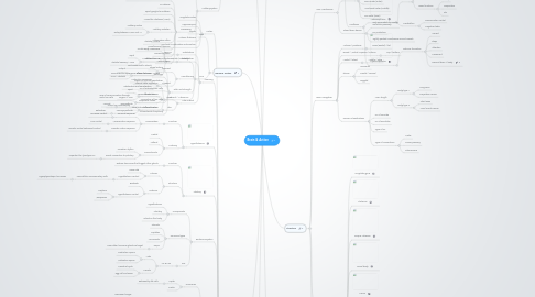
1. Nervous System
1.1. CNS
1.2. PNS
1.2.1. Somatic (voluntary)
1.2.1.1. Skeletal Muscle
1.2.1.2. Crude Body Awareness
1.2.1.3. Afferent nerves
1.2.1.3.1. Input
1.2.1.3.2. Skeletal Sensory > CNS
1.2.1.4. Efferent nerves = output
1.2.1.4.1. Output
1.2.1.4.2. CNS > Skeletal
1.2.2. Visceral / Autonomic
1.2.2.1. Internal state regulation
1.2.2.2. Afferent nerves
1.2.2.2.1. Input
1.2.2.2.2. Organs > CNS
1.2.2.3. Efferent nerves
1.2.2.3.1. Sympathetic
1.2.2.3.2. Para-sympathetic
2. Brain Measurement
2.1. Anatomy
2.1.1. CT Scan
2.1.1.1. Computed Tomography - X ray
2.1.2. MRI
2.1.2.1. Magnetic Resonance Imaging
2.1.2.2. More detailed than CT
2.1.2.3. 3D image
2.1.3. Cortical somatotopy
2.1.3.1. Mapping of cortex to stimuli
2.2. Function
2.2.1. PET
2.2.1.1. Positron emission tomography
2.2.1.2. Radioactive Tracer Introduced
2.2.1.2.1. Glucose linked
2.2.2. EEG
2.2.2.1. Electroencephalography
2.2.2.2. Electrical activity over scalp
2.2.3. fMRI
2.2.3.1. Functional MRI
2.2.3.2. Track blood flow / oxygen
2.2.4. Single-Cell
2.2.4.1. Microelectrode
2.3. Links
2.3.1. Neuroanatomic tracing techniques
2.3.1.1. Anterograde
2.3.1.1.1. Trace path of axons downstream
2.3.1.2. Retrograde
2.3.1.2.1. Trace upstream
3. Systems
3.1. Limbic
3.1.1. Cingulate cortex
3.1.2. Hippocampus
3.1.3. Fornix
3.1.4. Anterior thalamus
3.1.5. Involved in motivation and emotion
3.2. Learning
3.2.1. Habituation
3.2.1.1. Less NT
3.2.2. Sensitization
3.2.2.1. More stimulus = amplification
3.2.3. Conditioning
3.2.3.1. Operant
3.2.3.1.1. Not connected to stimuli
3.2.3.1.2. Connected to experience of own behavior
3.2.3.2. Classical
3.2.3.2.1. 2 stimuli 2 responses
3.2.3.2.2. Habituation and sensitization
3.2.4. Like vs want
3.2.4.1. Want
3.2.4.1.1. Desire for stimulus
3.2.4.1.2. Dopamine linked
3.2.4.2. Like
3.2.4.2.1. Taste/distaste for stimulus
3.3. Hypothalamus
3.3.1. Function
3.3.1.1. Homeostasis
3.3.1.1.1. Humoral response
3.3.1.1.2. Visceromotor response
3.3.1.1.3. Somatic motor response
3.3.2. Anatomy
3.3.2.1. Medial
3.3.2.2. Latteral
3.3.2.3. Perivertricular
3.3.2.3.1. Circadian rhythm
3.3.2.3.2. Direct connection to pituitary
3.4. Pituitary
3.4.1. Function
3.4.1.1. Release hormones that trigget other glands
3.4.2. Structure
3.4.2.1. Anterior
3.4.2.1.1. Nose side
3.4.2.1.2. Hypothalamic control
3.4.2.2. Posterior
3.4.2.2.1. Backside
3.4.2.2.2. Hypothalamic control
3.5. Endocrine system
3.5.1. Components
3.5.1.1. Hypothalamus
3.5.1.2. Pituitary
3.5.1.3. Glands in the body
3.5.2. Hormone types
3.5.2.1. Steroids
3.5.2.2. Peptides
3.5.2.3. Aminoacids
3.5.2.4. Tropic
3.5.2.4.1. Have other hormone glands as target
3.5.3. Sex
3.5.3.1. LH & FSH
3.5.3.1.1. Male
3.5.3.1.2. Female
3.6. Eating
3.6.1. Hormones
3.6.1.1. Leptin
3.6.1.1.1. Released by fat cells
3.6.1.2. Insulin
3.6.2. Brain
3.6.2.1. Hypothalamus
3.6.2.1.1. POMC Circuit
3.6.2.1.2. NPY Circuit
3.6.3. Short term regulation
3.6.3.1. Phases
3.6.3.1.1. Cephalic
3.6.3.1.2. Gastric
3.6.3.1.3. Substrate
3.6.3.2. Substances
3.6.3.2.1. CCK
3.6.3.2.2. Insulin
3.6.3.2.3. PYY
3.7. Motor control
3.7.1. Levels
3.7.1.1. High
3.7.1.1.1. Association area (goal)
3.7.1.1.2. Basal ganglia (strategy)
3.7.1.2. Medium
3.7.1.2.1. Cerebellum
3.7.1.2.2. Motor cortex
3.7.1.2.3. Smooth muscle movements
3.7.1.3. Low
3.7.1.3.1. Brain stem
3.7.1.3.2. Spinal cord
3.7.1.3.3. Actual muscle coordination
3.7.2. Pathways
3.7.2.1. Rubrospinal
3.7.2.1.1. Reflex movement
3.7.2.2. Ventromedial
3.7.2.2.1. Proximate joints
3.7.2.2.2. Bodyposition and balance
3.7.2.3. Lateral
3.7.2.3.1. Distal joints
3.7.2.3.2. Corticoreticolospinal tract
3.7.3. Areas
3.7.3.1. Cerebellum
3.7.3.1.1. Association cortex
3.7.3.1.2. Cerebellum
3.7.3.1.3. SMA
3.7.3.2. Basal Ganglia
3.7.3.2.1. Association cortex
3.7.3.2.2. Putamen
3.7.3.2.3. Globus pallidus
3.7.3.2.4. VLO
3.7.3.2.5. SMA
3.7.3.2.6. Motor tasks but also others
3.7.3.3. Somatosensory Cortex
3.7.3.3.1. Most in parietal lobe
3.7.3.3.2. Primary (S1) on postcentral gyrus
3.7.3.3.3. M1 is more sensitive to sensory input
3.8. Sleep
3.8.1. Regulation
3.8.1.1. By
3.8.1.1.1. Superchiasmic nucleus
3.8.1.1.2. Hypothalamus
3.8.1.1.3. Photoreceptors
3.8.1.2. Promoting factors
3.8.1.2.1. Adenosin
3.8.1.2.2. Melatonin
3.8.1.2.3. Muramyl
3.8.1.2.4. SCN
3.8.2. Phases
3.8.2.1. REM
3.8.2.1.1. High frequency
3.8.2.1.2. Low amplitude
3.8.2.2. N-REM
3.8.3. Brainwaves
3.8.3.1. Beta 14+
3.8.3.2. Alpha 8-13
3.8.3.2.1. Quiet waking
3.8.3.3. Theta 4-7
3.8.3.3.1. Some sleep states
3.8.3.4. Delta 4-
3.8.3.4.1. High amplitude deep sleep
4. Organs
4.1. Eye
4.1.1. Structure
4.1.1.1. General and cells
4.1.1.1.1. Cornea
4.1.1.1.2. Iris
4.1.1.1.3. Retina
4.1.1.1.4. Ciliary muscle
4.1.1.1.5. Fovea
4.1.1.1.6. Optic nerve
4.1.1.1.7. Lens
4.1.1.1.8. Zolune fibres
4.1.1.1.9. Sclera
4.1.1.1.10. Liquids
4.1.1.2. Layers
4.1.1.2.1. Ganglion layer
4.1.1.2.2. Inner plexiform
4.1.1.2.3. Inner nuclear
4.1.1.2.4. Outer plexiformm
4.1.1.2.5. Outer nuclear layer
4.1.1.2.6. Photoreceptor outer segments
4.1.1.2.7. Pigmented epithelium
4.1.2. Processing
4.1.2.1. Done by
4.1.2.1.1. LGN
4.1.2.1.2. V1
4.1.2.1.3. Hypothalamus
4.1.2.1.4. Midbrain
4.1.2.2. Visual hemifields
4.2. Auditory system
4.2.1. Structure
4.2.1.1. Inner ear
4.2.1.1.1. Oval window
4.2.1.1.2. Cochlea
4.2.1.1.3. Organ of Corti
4.2.1.1.4. Basilar membrane
4.2.1.2. Middle ear
4.2.1.2.1. Tympanic membrane
4.2.1.2.2. Ossicles
4.2.1.3. Outer ear
4.2.1.3.1. Pinna
4.2.1.3.2. Audatory canal
4.2.2. Processing
4.2.2.1. Depolarisation of hair cells in Organ or Corti
4.2.2.2. NT release
4.2.2.3. Spiral ganglia to midbrain
4.2.2.4. Areas like Thalamus (MGN)
4.2.2.5. Auditory radiation
4.2.2.5.1. Auditory cortex
4.2.2.5.2. Delay between MGN and A1
4.2.2.6. Attenuation reflex
4.2.2.7. Mono/binaural neurons
4.2.2.8. Horizontal plane
4.2.2.9. Vertical plane
4.2.3. Pitch and strength
4.2.3.1. Phase locking
4.2.3.2. Tonotopy
4.2.3.3. Depth of cochlea penetration
4.2.3.4. Nr of activated hair cells
4.2.3.5. Innervation of hair cells
4.2.3.5.1. 95% of communication through inner hair cells
4.2.3.5.2. Outer hair cells amplify
4.2.3.6. Characteristic frequency
5. Brain Structure
5.1. Lobes
5.1.1. Parietal Lobe
5.1.1.1. Positioning
5.1.1.2. Knowing where the body is
5.1.1.3. In the middle "Part"
5.1.2. Frontal Lobe
5.1.2.1. Reasoning
5.1.2.2. Remembering
5.1.2.3. Executive decision maker
5.1.2.4. In the front
5.1.3. Occipital Lobe
5.1.3.1. Contains visual cortex
5.1.3.2. Sensory information
5.1.3.3. Occi = eyes, at the back
5.1.4. Temporal Lobe
5.1.4.1. Auditory Cortex
5.1.4.2. Language
5.1.4.3. Time underlies everything = bottom
5.1.5. Not functional units on their own
5.2. Levels
5.2.1. Brainstem
5.2.1.1. Live
5.2.2. Medial Level
5.2.2.1. Diencephalon
5.2.2.2. limbic system
5.2.2.3. Food, Fight, Fuck
5.2.3. Cerebral Cortex
5.2.3.1. The Lobes
5.2.3.2. Insular Cortex
5.2.3.3. Cerebellum
5.2.3.4. Structure
5.2.3.4.1. Fissures
5.2.3.4.2. Sculi
5.2.3.4.3. Gyri
5.2.3.4.4. Cortex
5.3. Brain Division
5.3.1. Forebrain
5.3.1.1. Telencephalon / Cerebrum
5.3.1.1.1. Largest division
5.3.1.1.2. Voluntary movement
5.3.1.1.3. Interpret sensory input
5.3.1.1.4. Mediates complex cognitive processes
5.3.1.1.5. Divisions
5.3.1.2. Diencephalon
5.3.1.2.1. Thalamus
5.3.1.2.2. Hypothalamus
5.3.2. Midbrain
5.3.2.1. Mesencephalon
5.3.2.1.1. Tectum
5.3.2.1.2. Tegmentum
5.3.3. Hindbrain
5.3.3.1. Metencephalon
5.3.3.1.1. Pons
5.3.3.1.2. Cerebellum
5.3.3.2. Myelencephalon
5.3.3.2.1. Reticuar formation
6. Structure
6.1. CNS Membranes
6.1.1. Menenges
6.1.1.1. Cerebrospinal fluid
6.1.1.1.1. Subarachnoid space
6.1.1.1.2. Protects against mechanical schock
6.1.1.1.3. Produced in Choroid plexuses
6.1.1.2. Dura mater (outer)
6.1.1.3. Arachnoid mater (middle)
6.1.1.4. Pia mater (inner)
6.1.2. Blood brain barrier
6.1.2.1. Only permeable to smaller molecules passively
6.1.2.2. For protection
6.1.2.3. Tightly packed membranes around vessels
6.2. Brain Navigation
6.2.1. Anterior / posterior
6.2.1.1. Nose (Rostral) / tail
6.2.2. Dorsal / Ventral, Superior / inferior
6.2.2.1. Top / bottom
6.2.3. Medial / lateral
6.2.3.1. Middle / sides
6.2.4. Planes
6.2.4.1. Horizontal
6.2.4.2. Frontal / coronal
6.2.4.3. Saggital
6.2.5. Neuron Classifications
6.2.5.1. Axon length
6.2.5.1.1. Golgi type I
6.2.5.1.2. Golgi type II
6.2.5.2. Nr of Neurites
6.2.5.3. Nr of Dendrites
6.2.5.4. Type of NT
6.2.5.5. Type of connections
6.2.5.5.1. Motor
6.2.5.5.2. Primary sensory
6.2.5.5.3. Interneurons
6.3. Important parts orientation
6.3.1. Cingulate gyrus
6.3.2. Thalamus
6.3.3. Corpus calossum
6.3.4. Pineal body
6.3.5. Fornix
6.3.6. Hypothalamus
6.3.7. Pons
6.3.8. Cerebellum
6.3.9. Medulla
6.3.10. Basal Ganglia
6.3.10.1. Caudate nucleus
6.3.10.2. Putamen
6.3.10.3. Globus pallidus
6.3.10.4. Somatic NS
6.3.10.5. Movement coordination
6.4. Spinal Cord
6.4.1. Cervical
6.4.1.1. C1-C7
6.4.1.1.1. Neck
6.4.2. Thoraic
6.4.2.1. T1-T12
6.4.2.1.1. Attached to ribs
6.4.3. Lumbar
6.4.3.1. L1-L5
6.4.3.1.1. Lower back
6.4.4. Sacral
6.4.4.1. S1-S?
6.4.4.1.1. Pelvic area
7. Cellular
7.1. Nervous system cells
7.1.1. Neurons
7.1.1.1. Dendrite
7.1.1.1.1. Root like structure
7.1.1.2. Cell body
7.1.1.2.1. Golgi apparatus
7.1.1.2.2. Dendritic spinals
7.1.1.2.3. Free ribosome
7.1.1.2.4. Rough ER
7.1.1.2.5. Smooth ER
7.1.1.2.6. Lysosome
7.1.1.2.7. Axon hillock
7.1.1.2.8. Microtubules
7.1.1.2.9. Mitochondria
7.1.1.3. Myelin sheath
7.1.1.3.1. rolled up along axon
7.1.1.4. Nodes of Ranvier
7.1.1.4.1. Between myelin
7.1.1.5. Axon
7.1.1.5.1. the tail
7.1.1.6. Axon terminal
7.1.1.6.1. the end of the tail
7.1.2. Glial Cells
7.1.2.1. Nourishment and support
7.1.2.2. Astrocytes
7.1.2.2.1. Maintain extracellular state
7.1.2.2.2. Contain NT action
7.1.2.3. Myelinating Glia
7.1.2.3.1. Schwann cell
7.1.2.3.2. Oligodendrogia
7.2. Neuro transmitters
7.2.1. Synthesis
7.2.1.1. In Cell body
7.2.1.1.1. RER
7.2.1.1.2. Transport to axon terminal
7.2.1.1.3. Axoplasmic transport
7.2.1.2. Axon terminal
7.2.1.2.1. GABA
7.2.1.2.2. Amines
7.2.1.3. Outside cell
7.2.2. Types
7.2.2.1. Aminoacids
7.2.2.2. Amines
7.2.2.3. Peptides
7.3. Action potential
7.3.1. Rising phase
7.3.2. Depolarisation
7.3.3. Na influx
7.3.4. Fall phase
7.3.5. Repolarisation
7.3.6. K efflux
7.3.7. Hyperpolarisation
7.3.8. Resting potential
7.4. Synaptic transmission
7.4.1. Influx of ca++ causes exocytosis
7.4.2. Vesicle restored in endocytosis
7.4.3. EPSP
7.4.3.1. Excitatory post synaptic potential
7.4.4. IPSP
7.4.4.1. Inhibiting post synaptic potential
7.4.5. Receptors
7.4.5.1. Types
7.4.5.1.1. G-protein coupled
7.4.5.1.2. Autoreceptors
7.4.5.1.3. Ion channel
7.4.5.2. Binders
7.4.5.2.1. Substance
7.4.5.2.2. Agonist
7.4.5.2.3. Antagonist
7.4.6. Summation
7.4.6.1. Spatial
7.4.6.1.1. Signals from multiple neurons
7.4.6.2. Temporal
7.4.6.2.1. Higher frequency of excitement
