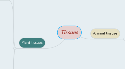
1. Plant tissues
1.1. Meristematic cells
1.1.1. Small
1.1.2. Thin walled
1.1.3. Cubical
1.1.4. Produce new cells by cell-division
1.1.5. Packed with protoplasm
1.1.6. Top tips of roots and stems , between the water and food conducting
1.2. Permanent tissues
1.2.1. Parenchyma cells
1.2.1.1. Most abundant
1.2.1.2. Alive
1.2.1.3. Lack secondary
1.2.1.4. Thin primary
1.2.1.5. Photosynthesis , storage
1.2.2. Don't become changed
1.2.3. Collenchyma cells
1.2.3.1. Unevenly thickened primary
1.2.3.2. Flexible support
1.2.3.3. Lack secondary
1.2.3.4. Alive
1.2.4. Sclerenchyma cells
1.2.4.1. Thick secondary
1.2.4.1.1. Lignin
1.2.4.2. Rigid support
1.2.4.3. Dead
1.2.4.4. Type of it
1.2.4.4.1. Fibers : long , thin , in boundles
1.2.4.4.2. Sclereids : shorter than fibers
1.2.5. Water conducting cells ( tracheids and vessel elements )
1.2.5.1. Thick secondary
1.2.5.2. Dead
1.2.5.3. Chains of tracheids and vessel elements form xylem
1.2.6. Food conducting cells ( sieve tube members )
1.2.6.1. No secondary
1.2.6.2. Alive , lack most organelles
1.2.6.3. Companion cells
1.2.6.3.1. Organelles
1.2.6.3.2. Control operation of sieve tube members
1.2.6.4. Chains of seive tube members separated by porous form phloem
1.3. Tissue ystems that make up the plant body
1.3.1. Dermal tissue : outer protective covering
1.3.1.1. Epidermis: layer of tightly packed cells
1.3.1.1.1. On top of it there is a waxy layer ( cuticle ) that reduces water loss
1.3.1.2. First line of defense against damage and infection
1.3.2. Vascular tissue : support and transport
1.3.2.1. Xylem / الخشب
1.3.2.1.1. Water ( H2O)
1.3.2.2. Phloem / اللحاء
1.3.2.2.1. Food
1.3.2.3. In bundles
1.3.3. Ground : bulk of the plant body , food production, storage , support
1.3.3.1. Between dermal and vascular tissue
1.3.3.2. Eudicot stem ground tissue is divided into pith and cortex
1.3.3.3. Mesophyll : leaf ground tissue
1.4. Structure that distinguish plant cells from animal cells
1.4.1. 1- chloroplasts
1.4.2. 2- A large fluid-filled vacuole
1.4.3. 3- A cell wall composed of cellulose
1.5. Plant cell walls
1.5.1. There is two walls
1.5.1.1. Primary cell walls - outer most layer
1.5.1.2. Secondary cell walls
1.5.1.2.1. Tough layer inside primary wall
1.5.2. Middle lamella : a sticky layer lies between adjacent plant cells
1.5.3. Plasmodesmata : opining in cell walls allow cells to communicate and exchange material
2. Animal tissues
2.1. - Epithelial tissue
2.1.1. Simple squamous epithelium
2.1.1.1. Air sacs of the lung
2.1.2. Simple cuboidal epithelium
2.1.2.1. Kidney
2.1.3. Simple columnar epithelium /
2.1.3.1. Intestine /
2.1.4. Pseudostratified ciliated columnar epithelium
2.1.4.1. Respiratory tract
2.1.5. Stratified squamous epithelium
2.1.5.1. Esophagus /
2.2. - Cconnective tissue
2.2.1. Loose connective tissue /
2.2.1.1. Under the skin
2.2.2. Fibrous connective tissue
2.2.2.1. Forming a tendon
2.2.3. Adipose tissue
2.2.4. Cartilage
2.2.4.1. At the end of a bone
2.2.5. Bone
2.2.6. Blood
2.3. - Muscle tissue
2.3.1. Skeletal muscle
2.3.1.1. Voluntary movements
2.3.2. Cardiac muscle /
2.3.2.1. Pumps blood
2.4. - Nervous tissue
2.4.1. Neurons
2.4.1.1. Carry signals
2.4.2. Supporting cells insulate axons and nourish neurons
2.4.3. Smooth muscle
2.4.3.1. Moves walls of internal organs
2.4.3.1.1. Intestines
