

1. Prostate Stercz
1.1. Inflammatory
1.1.1. Acute bacterial prostatitis
1.1.2. Chronic bacterial prostatitis
1.1.3. Chronic abacterial prostatitis/prostatodynia
1.2. Tumors
1.2.1. Nodular hyperplasia/Benign prostatic hyperplasia (BPH) Łagodny rozrost stercza
1.2.1.1. General Ogólne
1.2.1.1.1. Very common
1.2.1.2. Pathology
1.2.1.2.1. Nodular hyperplasia, stroma&glands Guzkowy rozrost
1.2.1.2.2. Transition zone Strefa przejsciowa
1.2.2. Carcinoma
1.2.2.1. General
1.2.2.1.1. Very common RISK FACTORS: AGE, GENETIC, ETHNICITY CLINICAL PRESENTATION: LATE DUE TO METASTASES
1.2.2.1.2. DRE/PSA Badanie per rectu/ Antygen sterczowy w
1.2.2.1.3. Latent vs clinically significant
1.2.2.2. Pathology
1.2.2.2.1. Adenocarcinoma
1.2.2.2.2. Peripheral zone
1.2.2.2.3. Precursors: HGPIN, Adenosis (AAH)
1.2.2.2.4. Spread
1.2.2.2.5. Role of pathologists
1.2.2.3. Biopsy, core bx
1.2.2.3.1. diagnosis
1.2.2.3.2. GS and tumor volume Wskażnik Gleasona i objętosć guza
1.3. Anatomy & histology
1.3.1. Untitled
1.3.1.1. Zonal anatomy
1.3.1.1.1. Zonal anatomy
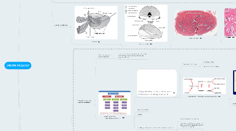
2. Testis & paratesticular tissues Jądro i tkanki okołojądrowe
2.1. Congenital anomalies Anomalie wrodzone
2.1.1. Testicular dysgenesis Dysgenezja jąder
2.1.1.1. abnormal embryonic development of gonads (hypoplastic or incompletely developed+ hypogonadism))
2.1.2. EMPTY SCROTUM BRAK JĄDRA W MOSZNIE
2.1.2.1. Cryptorchidism - undescended testis Wnętrostwo - niezstąpienie jądra
2.1.2.1.1. Treatment: orchiopexy
2.1.2.1.2. component of Testicular Dysgenesis Syndrome
2.1.2.2. Retractile testicle
2.1.2.3. Ectopy
2.1.2.4. Vanishing testes syndrome/testicular regression syndrome
2.1.2.4.1. At least 5% of empy scrotum
2.1.2.4.2. vascular accident/va during embry or fetal development leading to loss of initially normal testicle
2.1.2.4.3. phenotype depends on the time of va - variants from unilateral anorchia through ambiguous genitalia to female internal & external phenotype
2.1.2.5. Agenesis
2.1.2.6. PATHOLOGY depends on the type and includes:atrophy-> fibrous streak->complete lack of testicularstructures
2.2. Inflammatory
2.2.1. Epididymo-orchitis Zapalenie jądra i najądrza
2.2.1.1. Infectious (non-specific and STD)
2.2.1.2. Non-infectious (vasculitis, trauma, idiopathic)
2.2.2. Orchitis
2.2.2.1. Mumps
2.2.3. Epididymitis
2.2.3.1. Tuberculosis
2.3. Vascular
2.3.1. Torsion
2.3.1.1. anomalous testicular suspension defect
2.3.1.1.1. torsion
2.4. Germ cell tumors (GCTs) >95%
2.4.1. General
2.4.1.1. Most common cancer in young males
2.4.1.2. Aggresive but curable
2.4.1.3. AGE DIFFERENCES
2.4.1.4. Biomarkers: AFP, bHCG, LDH
2.4.2. Pathology
2.4.2.1. Pure (40%)
2.4.2.1.1. Seminoma (>90% of pure, 35-40% of all)
2.4.2.1.2. Non-seminomatous
2.4.2.2. Mixed most common (60%)
2.4.2.3. Precursor: IGCN
2.4.2.4. Spermatocytic seminoma
2.4.2.4.1. distinct entity, different molecular pathway, elderly
2.5. Tumors
2.5.1. Neoplasms
2.5.1.1. current view on the development of type II germ cell tumors
2.5.1.2. Sex cord stromal tumors
2.5.1.2.1. Leydig cell tumor (most common SCST)
2.5.1.3. Secondary
2.5.1.3.1. Lymphoma (most common testucular neo in >60 yo)
2.5.2. Celes and cysts of spermatic cord & adnexa
2.5.2.1. Hydrocele
2.5.2.2. Spermatocele
2.5.2.3. Varicocele
2.5.3. Differential diagnosis
2.5.3.1. Epidymoorchitis - swelling
2.5.3.2. Inguinal hernia
2.6. HYPOGONADISM (gonadal failure)
2.6.1. PRIMARY (hypergonadotropic)
2.6.1.1. genetic syndromes
2.6.1.2. cryptorchidism
2.6.1.3. vanishing testes syndrome
2.6.1.4. external insults (eg inflammation, ischaemia)
2.6.1.5. autoimmune
2.6.1.6. Sertoli cell only syndrome
2.6.2. SECONDARY (hypogonadotropic)
2.6.3. presentation depends on the age of development
2.7. Anatomy & histology
2.7.1. testis
2.7.1.1. seminiferous ducts
2.7.1.1.1. seminiferous ducts
3. Penis and urethra Prącie i cewka moczowa
3.1. Structural/Congenital Abnormalities
3.1.1. Abnormal urethral groove/canal
3.1.1.1. Hypospadias
3.1.1.2. Epispadias
3.1.2. Phimosis
3.1.2.1. Paraphimosis_(urologic_emergency)
3.1.3. Posterior_urethral_valve
3.2. Inflammatory
3.2.1. Balanoposthitis
3.2.1.1. infectious(STD&non-specific)
3.2.1.2. non-infectious(Lichen_sclerosus_et_atrophicus/Balanitis_xerotica_obliterans)
3.2.2. Urethritis
3.3. Neoplasms
3.3.1. Benign
3.3.1.1. Condylomata acuminata
3.3.1.1.1. Histopathology
3.3.2. SCC precursor_lesions
3.3.2.1. Clinical presentations
3.3.2.1.1. Bowen's disease
3.3.2.1.2. Erytroplasia of Queyrat
3.3.2.1.3. Bowenoid papulosis
3.3.2.1.4. Leucoplakia_on_shaft_or_prepuce
3.3.2.2. Histopathology(common)
3.3.2.2.1. Penile intraepithelial neoplasia PeIN 2/3 (HGSIL)
3.3.3. Malignancy
3.3.3.1. Squamous cell carcinoma(>90%)
3.3.3.1.1. 0,5% of male cancers,>50yo
3.3.3.1.2. 30-60% are HR-HPV-related
3.3.3.1.3. SCC
3.3.3.2. Basal cell and transitional cell carcinomas arising from the urethra
3.3.3.3. Rare:adenocarcinoma, melanoma and Kaposi sarcoma.
3.3.3.4. Extremely rare:Metastases
3.3.3.4.1. 70% of lesions are of pelvic origin (from genitourinary or recto-sigmoid primary tumors)
4. SURGICAL CONDITIONS OF THE KIDNEYS, URETERS & URINARY BLADDER Schorzenia urologiczne nerek,moczowdów i pęcherza moczowego
4.1. CONGENITAL & CYSTIC DISEASES CHOROBY WRODZONE I TORBIELE
4.1.1. CONGENITAL MALFORMATIONS
4.1.1.1. URINARY BLADDER
4.1.1.1.1. AGENESIS
4.1.1.1.2. EXSTROPHY
4.1.1.2. KIDNEY
4.1.1.2.1. AMOUNT
4.1.1.2.2. POSITION
4.1.1.2.3. DIFFERENTIATION
4.1.2. CYSTIC DISEASES
4.1.2.1. CONGENITAL & HEREDITARY WRODZONE I DZIEDZICZNE
4.1.2.1.1. CYSTIC DYSPLASIA
4.1.2.1.2. POLYCYSTIC KIDNEY DISEASES ZESPÓŁWIELOTORBIELOWATOSCI NEREK
4.1.2.1.3. cystic diseases involving medulla
4.1.2.2. AQUIRED WRODZONE
4.1.2.2.1. SIMPLE CYSTS
4.1.2.2.2. AQUIRED/DIALYSIS ASSOCIATED CYSTIC DISEASE
4.1.2.3. CYSTIC FORMS OF NEOPLASMS
4.2. OBTURACJA DRÓG MOCZOWYCH
4.3. TUMORS OF THE UT
4.3.1. KIDNEY NEOPLASMS
4.3.1.1. MALIGNANT
4.3.1.1.1. RENAL CELL CARCINOMA (RCC)
4.3.1.1.2. UROTHELIAL CA
4.3.1.1.3. NEO PEDIATRIC
4.3.1.2. BENIGN
4.3.1.2.1. CORTICAL ADENOMA
4.3.1.2.2. ONCOCYTOMA
4.3.1.2.3. ANGIOMYOLIPOMA
4.3.2. URINARY BLADDER TUMORS
4.3.2.1. GENERAL OGÓLNE
4.3.2.1.1. PEAK 65 yo, M:F 3:1
4.3.2.1.2. almost all sporadic; MULTIFOCALITY
4.3.2.1.3. RISK FACTORS
4.3.2.2. CLINICS PREZENTACJA KLINICZNA
4.3.2.2.1. painless hematuria bezbolesny krwiomocz
4.3.2.2.2. UT obstruction obturacja dróg moczowych
4.3.2.3. HISTOPATOLOGY
4.3.2.3.1. UROTHELIAL NEOPLASMS (>90%)
4.3.2.3.2. NON-UROTHELIAL CARCINOMA
4.3.2.4. STAGING
4.3.2.4.1. VARIOUS LEVELS OF INVASION