
1. v IIII
2. Gametogenesis
2.1. Epiblast Cell Line
2.1.1. Primordial Germ Cells (PGCs) - gamete precursors
2.1.1.1. Migrate to yolk sac (PGCs visibile here at 4th week)
2.1.1.2. Migrate from yolk sac through connecting stalk to dorsal body wall (high on posterior abdominal wall) during 4th to 6th weeks
2.1.1.2.1. Undergo rapid mitosis during migration (billions of PGCs)
2.1.1.3. Populate developing gonads
2.1.1.3.1. develop from vertically oriented mass of mesoderm called urogenital ridge
2.1.1.3.2. Upon arrival, PGCs induce formation of follicles in ovary and Sertoli Cells in testis
2.1.1.4. Spermatogenesis (continuous from puberty until death)
2.1.1.4.1. upon arrival to testis, PGCs go dormant and remain dormant at birth and until puberty
2.1.1.4.2. Puberty: PGCs differentiate under influence of endocrine system
2.1.1.5. Oogenesis
2.1.1.5.1. upon arrival to developing gonad, PGCs immediately begin differentiation into oogonia - rapid mitotic division (by 5th month, ~7 mil)
3. Ovulation
3.1. Menstrual Cycle
3.1.1. Day 1-14
3.1.1.1. Primordial follicle houses primary oocyte in 1st arrested state
3.1.1.2. Primordial follicle becomes primary follicle, adding a few outer cell layers (zona pellucida, corona radiata)
3.1.1.2.1. Grows into secondary follicle (larger, contains fluid filled antrum)
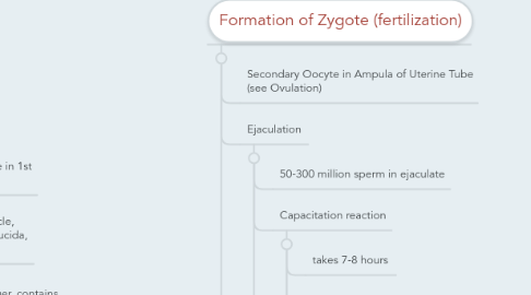
4. Formation of Zygote (fertilization)
4.1. Secondary Oocyte in Ampula of Uterine Tube (see Ovulation)
4.2. Ejaculation
4.2.1. 50-300 million sperm in ejaculate
4.2.2. Capacitation reaction
4.2.2.1. takes 7-8 hours
4.2.2.2. occurs while spermatozoa are traveling through uterine tube
4.2.2.3. rearranges proteins on acrosome cap of spermatozoa, rendering sperm capable of penetrating through corona radiata
4.2.3. 300-400 reach sec. oocyte
4.3. Spermatozoa line up around outer perimeter of sec. oocyte
4.3.1. Thanks to capacitation reaction, hundreds of sperm bore through corona radiata and attach to outer surface of zona pellucida
4.3.1.1. When head of sperm contacts zona pellucida, acrosome reaction occurs
4.3.1.1.1. this reaction opens pores on the head of the sperm for release of proteolytic enzymes that bore through zona pellucida
5. Development: Week 1
5.1. Days 1-4
5.1.1. Formation of diploid zygote
5.1.2. Fusion of pronuclei induces cleavage
5.1.2.1. Cleavage: a series of mitotic cell divisions
5.1.3. Zona Pellucida prevents cells from increasing in mass as well as preventing developing embryo from implanting uterine tube
5.2. Day 5
5.2.1. Morula (16-32 cells) stage/ endpoint of cleavage
5.2.1.1. morula is moved through uterine tube via movement of cilia in uterine tube, fluid movement, and muscular contraction
5.2.2. Zona Pellucida begins to break down
5.2.3. Cells of morula are reorganized to form Blastocyst
5.2.3.1. gives rise to first cell lines of development
5.2.3.2. Some cells are pushed to one pole, forming homogenous mass of cells called embryoblast
5.2.3.2.1. embryoblast gives rise to embryo proper (pleuripotent embryonic stem cells)
5.2.3.3. other cells form shell-like perimeter called trophoblast
5.2.3.3.1. forms placenta
5.2.3.4. blastocyst cavity (fluid filled) develops in blastocyst
5.3. Days 5-6
5.3.1. Blastocyst enters uterine cavity
5.3.2. floats freely for ~1 day
5.4. Day 6-7
5.4.1. blastocyst adheres to uterine lining
5.4.1.1. Embryoblast attaches to uterine wall first
5.4.2. Trophoblast cells differentiate into cytotrophoblasts and syncytiotrophoblasts (can undergo mitotic division in week 1 ONLY).
5.4.2.1. cytotrophoblasts occupy same position as old trophoblast layer, while syncytiotrophoblasts push in front of embryoblast and forms finger-like projections that release enzymes that allow embryo to erode mother's uterine lining during early stages of implantation
6. Development: Week 2
6.1. Day 8
6.1.1. Embryoblast reorganizes into bilaminar disc
6.1.1.1. Bilaminar disc: two distinct cell layers
6.1.1.1.1. dorsal surface: epiblast cells
6.1.1.1.2. ventral surface: hypoblast cells
6.1.2. Syncytiotrophoblasts erode further into uterine wall
6.1.2.1. cells do not have complete cell membranes, incapable of mitotic division
6.1.3. As cytotrophoblast cells divide, they push into mass of syncytiotrophoblast cells, eventually losing cell membranes and becoming part of syncytial mass
6.2. Days 9-10
6.2.1. Second fluid filled space called amnion forms
6.2.1.1. fills with amniotic fluid secreted by epiblast cells
6.2.1.2. completely lined by epiblast cells, epiblast cells forming roof of amnion known as amnioblasts
6.2.2. Hypoblast cells are metabolically active and send out a layer of cells that line the inner surface of the trophoblast surrounding blastocyst cavity
6.2.2.1. called Heuser's exocoelomic membrane
6.2.3. Old blastocyst cavity is now known as primary yolk sac
6.2.3.1. bounded by Heuser's Membrane (floor) and Hypoblast (ceiling)
6.2.4. Syncytiotrophoblast cells have penetrated uterine glands and materal bl vessels in uterine wall.
6.2.4.1. Uterine glands: supply glycogen, important nutrient for embryo
6.2.4.2. bl vessels: supply embryo with oxygenated bl
6.2.5. as syncytiotrophoblasts erode multiple maternal bl vessels, lacunar networks develop. Comprises rudimentary placental circulation. Diffusion suffices until placenta develops.
6.3. Days 11-12
6.3.1. Heuser's membrane lays down layer between the membrane and trophoblast layer = extraembryonic mesoderm
6.3.1.1. important for placental and umbilical cord
6.3.2. extraembryonic mesoderm develops vacuoles which coalesce to form chorionic space, dividing this mesoderm into inner and outer layers
6.3.2.1. Inner layer: visceral, extraembryonic splanchnic mesoderm
6.3.2.2. outer layer: extraembryonic somatic mesoderm
6.4. Days 13-14
6.4.1. Cells come off the hypoblast layer and push the primary yolk sac away from the embryo to the distal edge of chorion where it is discarded
6.4.2. Forms definitive yolk sac
6.4.2.1. houses PGCs early in development
6.4.2.2. early hematopoiesis
6.4.3. Connecting stalk forms in region dorsal to amnion, where layers of extraembryonic mesoderm are adjacent to one another. This will give rise to umbilical cord.
6.4.4. cytotrophoblasts project out into syncytial mass, forming columns of cells called primary villi
6.4.4.1. villi will eventually contain fetal vascular system
6.4.4.2. Physical proximity of maternal bl flow in lacunae and fetal blood flow in villi is basis for diffusion of O2 and nutrients in early weeks of development
6.4.5. Thickening at one end of bilaminar disc, known as prechordal plate, is future mouth and defines cranial and caudal ends of developing embryo
6.5. Syncytiotrophoblasts secrete hCG
6.5.1. Maintains corpus luteum in ovary after ovulation and through first trimester, allowing for production of estrogen and progesterone
6.5.2. Prevents menstrual cycle at day 28
7. Development: Week 3
7.1. Days 15-18
7.1.1. Gastrulation: migration of epiblast cells through primitive streak
7.1.1.1. Primitive streak forms along dorsal cranial-caudal axis on caudal half (demarcated by site of cloacal membrane (site of future anus)
7.1.1.1.1. epiblast cells along primitive streak harden, forming an elevated ridge. Center of ridge sinks down to form the streak, or groove.
7.1.1.1.2. cranial end of streak is called primitive node
7.1.1.2. Epiblast cells migrate through primitive streak to form trilaminar disc
7.1.1.2.1. 1) Cells migrate ventrally and replace hypoblast layer of old bilaminar disc. Hypoblast cells pushed laterally and then removed. Forms innermost germ cell layer - Endoderm
7.1.1.2.2. 2) epiblast cells fill in space between endoderm and epiblast layers, forming middle germ layer - Mesoderm
7.1.1.2.3. 3)last group of epiblast cells do not migrate through streak. They remain on dorsal surface of disc and form outermost germ cell layer - Ectoderm
7.1.1.3. After trilaminar disc is formed, other epiblast cells form a vertically oriented rod of tissue in midline of mesoderm layer = notochord (epiblast cells grow cranially from primitive node to from notochord)
7.1.1.3.1. Notochord extends from oropharyngeal membrane to cloacal membrane
7.1.1.4. Primitive streak regresses in caudal direction and is reabsorbed into caudal end of disc