

1. N. Opthalamicus
1.1. **rr. tentorius ** Sinus cavernosus
1.1.1. Meninges
1.2. Posterior to FOS divdes into 3 main branches
1.2.1. n. nasociliaris
1.2.1.1. through anulus tendineus communis
1.2.1.1.1. n. ethmoidalis ant
1.2.1.1.2. n. ethmoidalis post.
1.2.1.1.3. nn. ciliares breves
1.2.1.1.4. nn. ciliares longi
1.2.1.1.5. TERMINAL n.infratrochlearis
1.2.2. n.frontalis
1.2.2.1. runs along the orbital roof
1.2.2.1.1. supratrochlear & supraorbital nerves
1.2.3. n. lacrimalis
1.2.3.1. runs along the lateral orbital wall to
1.2.3.1.1. the lacrimal gland, lateral eye angle & upper eyelid
1.2.3.2. Postganglionic GVE (PS) fibers from ***ganglion pterygopalatinum* ** (N.facialis)
1.2.3.2.1. Lacrimal gland
1.2.3.3. R. communicans cum nervo zygomatico
1.3. terminal branches **rr. nasales externi**
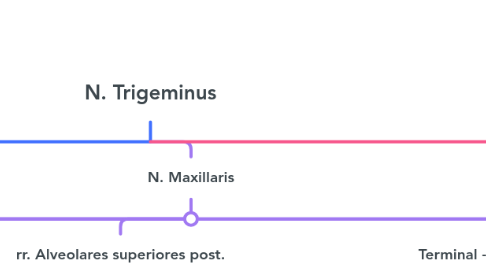
2. N. Maxillaris
2.1. R. meningeus (intracranially)
2.1.1. dura of the middle cranial fossa
2.2. N. Zygomaticus
2.2.1. • originates in fossa pterygopalatina • fissura orbitalis inferior --> orbita • there it gives off:
2.2.1.1. brings postganglionic GVE (PS) fibers from ganglion pterygopalatinum (N. FACIALIS) in orbita sends to n. lacrimalis for ps innervation of glandula lacrimalis
2.2.1.2. r. zygomaticofacial
2.2.1.2.1. Lateral eye angle
2.2.1.3. r. zygomaticotemporal
2.2.1.3.1. Skin of temple
2.3. rr. ganglionares ad ganglion pp
2.3.1. enter ganglion pterygopalatinum where they join GVE (PS) fibers fom n. facialis
2.4. rr. Alveolares superiores post.
2.4.1. foramina alveolaria
2.4.1.1. sinus maxillaris, upper molars & their gingiva
2.5. Terminal - n. infraorbitalis
2.5.1. passes within the orbital floor & exits through foramen infraorbitale
2.5.1.1. the skin of: • Upper lip / cheek region • Conjunctiva of lower eyelid • Lateral area of nasal wings
2.5.2. COLLATERAL r. alveolaris superior med + ant
2.5.2.1. maxillary sinus.
2.5.2.2. Together with the posterior rami they form PLEXUS DENTALIS SUPERIOR
2.5.2.2.1. upper teeth and corresponding gingiva
2.6. branches of g.pterygopalatinum
2.6.1. 1. GSA fibers (from n. maxillaris) 2. GVE fibers: post. ganglionic • PARASYMPATHETIC • SYMPATHETIC
2.6.1.1. 1. RR. NASALES SUPERIORES POSTERIORES LAT. & MED.
2.6.1.1.1. foramen sphenopalatinum
2.6.1.2. 2. N. PALATINUS MAJOR
2.6.1.2.1. canalis palatinus major
2.6.1.3. 3. NN. PALATINI MINORES
2.6.1.3.1. canalis palatinus minor
3. N. Mandibularis
3.1. r. meningeus
3.1.1. foramen spinosum
3.2. Ant division. N.masticatorius
3.2.1. Mainly SVE for mastication muscles
3.2.1.1. m.temporalis, m.masseter, mm. pterygoidei, but also for m. tensor veli palatini & m. tensor tympani
3.2.2. N. BUCCALIS - only afferent
3.2.2.1. for the buccal mucosa and skin
3.3. Post. Division - mixed fibres
3.3.1. N. aurioculotemporalis
3.3.1.1. Loops around a.meningea media
3.3.1.1.1. **GSA** - skin of the ear, ear drum & temple
3.3.1.1.2. **GVE(PS)PG** fibres from **GANGLION OTICUM (N.IX) ** for glandula parotidea.
3.3.2. N.alveolaris inferior
3.3.2.1. passes between pterygoid muscles --> canalis mandibulae
3.3.2.1.1. teeth of the lower jaw and corresponding gingiva (GSA fibers)
3.3.2.2. before foramen mandibule gives off
3.3.2.2.1. N.MYLOHYOIDEUS
3.3.2.3. foramen mentale
3.3.2.3.1. N. MENTALIS
3.3.3. N.lingualis
3.3.3.1. runs medially of n. alveolaris inferior & reaches the root of the tongue
3.3.3.1.1. with ITS own fibers (GSA) innervates
3.3.3.2. **Preganglionic GVE (PS)** fibers from **Nucleus salivatorius sup.** of **N. facialis.**
3.3.3.2.1. Via Chorda Tympani pass to N.lingualis in fossa infra temporalis
3.3.3.3. • SVA (taste) from taste buds at back & tip of tongue travel along lingual nerve (have nothing with ganglion submandibulare)
3.3.3.3.1. then along the chorda tympany