

1. Day 1
1.1. Fertilization
1.1.1. Sperm enters secondary oocyte
1.1.1.1. Corona radiata barrier
1.1.1.1.1. ZP3
1.1.1.2. Binding to ZP and acrosomal reaction
1.1.1.2.1. Acrosin
1.1.1.2.2. Integrins on oocytes
1.1.1.2.3. disintigrins on sperm
1.1.1.3. Fusion of sperm to oocyte
1.1.1.3.1. Permeability change of ZP
1.1.2. Oocyte completes meiosis II
1.1.2.1. Male and female pronucleus form
1.1.3. Initiation of cleavage
1.1.3.1. Two cells - 30 hours
1.1.3.2. Four cell blastomeres - 40 hours
2. Day 3
2.1. Morula
2.1.1. Compaction of embryoblast
2.1.1.1. 8-16 cells
3. Day 4
3.1. Early Blastocyst
3.1.1. Embryoblast
3.1.2. Trophoblast
3.2. Hatches from the ZP
4. Day 5
4.1. Late Blastocyst
4.1.1. Embryoblast begins to differentiate
4.1.1.1. Hypoblast
4.1.1.2. Epiblast
5. Day 6
5.1. Implantation
5.1.1. Endometrial cells of mother
5.1.1.1. Pinapodes
5.1.1.1.1. Leukemia inhibitory factor (LIF)
5.1.2. Cytotrophoblast
5.1.2.1. expresses the LIF receptor
5.1.3. Blastocyst adheres to endometrium
5.1.3.1. Starts producing own LIF
5.1.4. Endometrium produces additional attachment and tropic factors
5.1.4.1. Glycoprotein 130
6. Day 8
6.1. Bilaminar Disc
6.1.1. Embryoblast
6.1.1.1. Layer of columnar Epiblast cells
6.1.1.2. Layer of cuboidal Hypoblast cells
6.1.2. Trophoblast
6.1.2.1. Syncytiotrophoblast
6.1.2.1.1. Interleukin-1
6.1.2.2. Cytotrophoblast
7. Day 9
7.1. Lacunar Stage
7.1.1. Vacuoles appear in syncytium
7.1.1.1. Vacuoles fuse, creating lacunae
7.2. The blastocyst is more deeply embedded in the endometrium; penetration defect in the surface epithelium is closed by a fibrin coagulum
7.3. Epiblast
7.3.1. migrate and form amnioblasts
7.4. Hypoblast
7.4.1. Migrate and attach to cytotrophoblast cells
7.4.1.1. Line blastocyst cavity
7.4.1.2. Forms exocoelomic membrane
7.5. Blastocyst cavity becomes primitive yolk sac
8. Day 10-11
8.1. Blastocyst completely embedded in endometrium stroma
8.1.1. now produces a slight protrusion into the lumen of the uterus
8.2. surface epithelium almost entirely covers the original defect in the uterine wall
8.3. Embryonic Pole
8.3.1. Trophoblast characterized by lacunae in syncytium
8.3.1.1. Form intercommunicating network
8.4. Aembryonic Pole
8.4.1. Trophoblast still consists of mainly cytotrophoblastic cells
8.5. Syncytiotrophoblast penetrates deeper into the stroma
8.5.1. Erosion of maternal capillary endothelial lining
8.5.1.1. Formation of sinuses
8.5.1.1.1. Becomes part of lacunae
8.5.1.1.2. Establishment of uretoplacental circulation
9. Day 12
9.1. Extraembryonic mesoderm forms
9.1.1. Derived from yolk sac cells
9.1.2. Fine, loose connective tissue
9.1.3. Between the inner surface of the cytotrophoblast and the outer surface of the exocoelomic cavity
9.1.4. FIlls all of the space between the trophoblast externally and the amnion and exocoelomic membrane internally
9.1.5. large cavities develop, combine, and soon form extraembryonic cavity
9.1.5.1. Also called chorionic cavity
9.1.5.2. This space surrounds the primitive yolk sac and amniotic cavity
9.1.5.3. This space will later be eradicated by amniotic cavity growth due to the fusion of amnion and chorion to form anmniochorionic membrane
9.1.5.3.1. the amniochorionic membrane is what ruptures during labor
9.1.6. Extraembryonic somatic mesoderm
9.1.6.1. Lines the cytotrophoblast and amnion
9.1.7. Extraembryonic splanchnic mesoderm
9.1.7.1. Lines the yolk sac
9.2. Growth of bilaminar disc slower at this stage than trophoblast
9.2.1. Decidua reaction
9.2.1.1. Decidual cells degenerate adjacent to the syncytiotrophoblast and provide nutrition
9.3. hCG is secreted by syncytiotrophoblast
10. Day 13
10.1. Uretoplacental circulation begins
10.2. surface defect in the endometrium has usually healed
10.3. Bleeding may occur at this stage
10.3.1. It may be mistaken for menstruation
10.4. Primary villi forms
10.4.1. This (over time) will begin to protrude outward all the way to the uterine tissue over the syncytiotrophoblast
10.4.2. Will continue until differentiation and formation of villus the 4th week
11. Day 14
11.1. Embryonic Disc
11.2. Enough hCG for a pregnancy test
11.3. Bilaminar disc splitting identical twins at this stage
11.3.1. occurs just before appearance of primitive streak
11.3.2. Extremely rare
11.3.3. Will form identical twins in the same placenta with common chorionic and amniotic sacs
12. Day 15
12.1. Primitive streak begins to form
12.1.1. Marks the beginning of gastrulation
12.1.2. Primitive groove
12.1.3. Primitive node
12.1.4. Primitive pit
12.2. Laterally established
12.2.1. Streak is initiated and maintained by epiblast cells
12.2.1.1. Nodal
12.2.1.1.1. upregulates genes responsible for dorsal and ventral mesoderm
12.2.1.1.2. accumulates on left side near the node to establish left and right sidedness
12.2.1.2. epithelial-mesenchymal transformation
12.2.1.2.1. ingression of cells between the epiblast and hypoblast
12.2.1.2.2. This takes place days 16-17 to form trilaminar disc
13. Day 16
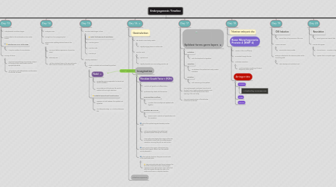
13.1. Gastrulation
13.1.1. The streak is now clearly visable
13.1.1.1. Slightly bulging lesions on either side
13.1.2. Primitive node
13.1.2.1. cephalic end
13.1.2.2. Slightly elevated area surrounding primitive pit
13.1.3. Invagination
13.1.3.1. Fibroblast Growth Factor 8 (FGF8)
13.1.3.1.1. Controls cell growth and differentiation
13.1.3.1.2. Synthesized by streak cells themselves
13.1.3.1.3. Downregulates E-cadherin
13.1.3.1.4. Regulates BRACHYURY
13.1.3.2. Cells of the epiblast migrate towards primitive streak
13.1.3.2.1. Cells moving between the epiblast and hypoblast spread laterally and cranially
13.1.3.2.2. These cells move beyond the margin of the disc and establish contact with the extraembryonic mesoderm covering the yolk sac and amnion
13.1.3.3. Upon arrival in the region of the streak, they become flask-shaped, detach from the epiblast, and slip beneath it
13.1.3.4. In the cephalic direction, they pass on each side of the prechordal plate.
13.1.3.4.1. The prechordal plate itself forms between the tip of the notochord and the oropharyngeal membrane and is derived from some of the first cells that migrate through the node in the midline and move in a cephalic direction
13.2. allantois appears
14. Day 17
14.1. Epiblast forms germ layers
14.1.1. Endoderm
14.1.1.1. Cells that displace the hypoblast
14.1.2. Mesoderm
14.1.2.1. Lies between the epiblast and newly created endoderm
14.1.3. Ectoderm
14.1.3.1. Cells remaining in the epiblast
14.2. The oropharyngeal membrane (cranial end of the disc) forms - tightly adherent ectoderm and endoderm cells that represents the future opening of the oral cavity.
14.3. The most cranial portion of the definitive notochord has formed
15. Day 18
15.1. Trilaminar embryonic disc
15.2. Bone Morphogenetic Protein 4 (BMP 4)
15.2.1. Member of the TGF-β family
15.2.2. Is secreted through the disc
15.2.3. Ventralizes mesoderm
15.2.3.1. Would ventralize everything if it wasn't stopped by other genes
15.2.4. Antagonists:
15.2.4.1. CHORDIN
15.2.4.1.1. Activated by GOOSECOID
15.2.4.2. noggin
15.2.4.3. follistatin
16. Day 19
16.1. CNS Induction
16.1.1. Neural tube is the precursor of the CNS
16.2. Amnion removed
16.2.1. Neural plate clearly visable
16.3. Embryo is attached to the chorionic plate via the connecting stalk
16.3.1. Later develops into umbilical cord
17. Day 20
17.1. Neurulation
17.1.1. Neural groove and neural folds
17.2. Somites first appear
17.2.1. This continues ~ 3 somites a day
17.2.2. Appears first in occipital region
18. Day 21
18.1. Transverse section
18.2. Beginning of third week of development
19. Day 22
19.1. Neural Tube closure begins
20. Day 23
20.1. Neural tube zippers
20.2. Embryo will be straight and slightly curved
20.2.1. still open on caudal end
21. Day 24-25
21.1. Villus formation
21.1.1. Mesodermal cells in villus core differentiate into blood cells and smaller blood cells
21.1.1.1. Forms the villus capillary system
21.1.2. Villus is now a tertiary villus or definitive placental villus