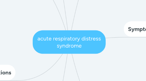
1. Causes
1.1. Sepsis. The most common cause of ARDS is sepsis, a serious and widespread infection of the bloodstream.
1.2. Inhalation of harmful substances. Breathing high concentrations of smoke or chemical fumes can result in ARDS, as can inhaling (aspirating) vomit or near-drowning episodes.
1.3. Severe pneumonia. Severe cases of pneumonia usually affect all five lobes of the lungs.
1.4. Head, chest or other major injury. Accidents, such as falls or car crashes, can directly damage the lungs or the portion of the brain that controls breathing.
1.5. Others. Pancreatitis (inflammation of the pancreas), massive blood transfusions and burns.
2. Risk factors
2.1. Most people who develop ARDS are already hospitalized for another condition, and many are critically ill. You're especially at risk if you have a widespread infection in your bloodstream (sepsis). People who have a history of chronic alcoholism are at higher risk of developing ARDS. They're also more likely to die of ARDS.
3. Complications
3.1. Blood clots. Lying still in the hospital while you're on a ventilator can increase your risk of developing blood clots, particularly in the deep veins in your legs. If a clot forms in your leg, a portion of it can break off and travel to one or both of your lungs (pulmonary embolism) — where it blocks blood flow.
3.2. Collapsed lung (pneumothorax). In most ARDS cases, a breathing machine called a ventilator is used to increase oxygen in the body and force fluid out of the lungs. However, the pressure and air volume of the ventilator can force gas to go through a small hole in the very outside of a lung and cause that lung to collapse.
3.3. Infections. Because the ventilator is attached directly to a tube inserted in your windpipe, this makes it much easier for germs to infect and further injure your lungs.
3.4. Scarring (pulmonary fibrosis). Scarring and thickening of the tissue between the air sacs can occur within a few weeks of the onset of ARDS. This stiffens your lungs, making it even more difficult for oxygen to flow from the air sacs into your bloodstream.
4. Treatment
4.1. Oxygen
4.1.1. Supplemental oxygen. For milder symptoms or as a temporary measure, oxygen may be delivered through a mask that fits tightly over your nose and mouth. Mechanical ventilation. Most people with ARDS will need the help of a machine to breathe. A mechanical ventilator pushes air into your lungs and forces some of the fluid out of the air sacs.
4.2. Fluids
4.2.1. Carefully managing the amount of intravenous fluids is crucial. Too much fluid can increase fluid buildup in the lungs. Too little fluid can put a strain on your heart and other organs and lead to shock.
4.3. Medication
4.3.1. People with ARDS usually are given medication to: Prevent and treat infections Relieve pain and discomfort Prevent blood clots in the legs and lungs Minimize gastric reflux Sedate
5. Overview
5.1. Acute respiratory distress syndrome (ARDS) occurs when fluid builds up in the tiny, elastic air sacs (alveoli) in your lungs. The fluid keeps your lungs from filling with enough air, which means less oxygen reaches your bloodstream. This deprives your organs of the oxygen they need to function.
5.2. ARDS typically occurs in people who are already critically ill or who have significant injuries. Severe shortness of breath — the main symptom of ARDS — usually develops within a few hours to a few days after the precipitating injury or infection.
5.3. Many people who develop ARDS don't survive. The risk of death increases with age and severity of illness. Of the people who do survive ARDS, some recover completely while others experience lasting damage to their lungs.
6. Symptoms
6.1. Severe shortness of breath
6.2. Labored and unusually rapid breathing
6.3. Low blood pressure
6.4. Confusion and extreme tiredness
7. Diagnosis
7.1. There's no specific test to identify ARDS. The diagnosis is based on the physical exam, chest X-ray and oxygen levels. It's also important to rule out other diseases and conditions — for example, certain heart problems — that can produce similar symptoms.
7.1.1. Chest X-ray. A chest X-ray can reveal which parts of your lungs and how much of the lungs have fluid in them and whether your heart is enlarged.
7.1.2. Computerized tomography (CT). A CT scan combines X-ray images taken from many different directions into cross-sectional views of internal organs. CT scans can provide detailed information about the structures within the heart and lungs.
7.2. Lab tests
7.2.1. A test using blood from an artery in your wrist can measure your oxygen level. Other types of blood tests can check for signs of infection or anemia. If your doctor suspects that you have a lung infection, secretions from your airway may be tested to determine the cause of the infection.
7.3. Heart tests
7.3.1. Electrocardiogram. This painless test tracks the electrical activity in your heart. It involves attaching several wired sensors to your body.
7.3.2. Echocardiogram. A sonogram of the heart, this test can reveal problems with the structures and the function of your heart.

